Family Seguenziidae Verrill, 1884
Seguenzia sp.
(Figs. 66 a-f, 67 a-h)
Protoconch: Protoconch well-inflated, conispiral. Lateral pouch smooth, without disjunct extension (Figs. 66 b-d). Suture of protoconch very clearly demarcated. Growth direction of protoconch not hyperstrophic, but orthostrophic ventrally. Surface homogeneously covered with somewhat spirally arranged, irregular elongated deposits (Fig. 66 f). Protoconch-teleoconch boundary gently thickened by broad apertural ridge (Fig, 66 e).

External Anatomy: Shell and animal trochiform. Mantle with paired (inhalant and exhalant) slits located in corresponding positions of sinuses of shell aperture. Mantle margin mamillate with microtentacles having ciliary corona (Fig. 67 a). Long pallial tentacles absent. Attachment of shell muscle single, slightly coiling along columella of shell. Muscle not divided into bundles.

Head with elongated snout and pair of papillate cephalic tentacles (ct) (Fig. 67 b). Cephalic lappets and neck lobes absent. Snout simple. Outer lip of mouth without oral lappets. Cephalic tentacles thicker than epipodial tentacles, with ciliated microtentacles shortened near bases but becoming longer toward tips of cephalic tentacles. Subocular peduncle and "accessory cephalic process" (Quinn, 1983) arising from outer base of right cephalic tentacle and from right side of neck region, respectively.
Foot sole not divided by mid-ventral cleft. Ventral surface of foot densely ciliated. Opening of pedal gland present anteriorly. Epipodium with three pairs of papillate epipodial tentacles (Fig. 67 b) with microtentacles on which ciliary crowns occur (Fig, 67 c). Paucispiral corneous operculum present on epipodium.
Pallial Complex: Pallial cavity very deep, occupying more than one-half whorl, containing left ctenidium, anus, and left hypobranchial gland. Ctenidium completely monopectinate, consisting of nearly 30 ctenidial filaments attached to mantle (Fig. 67 d). Ctenidial filaments heavily ciliated. Presence of bursicles suggested by ciliated groove (Fig. 67 d). Presence of osphradium not verified. Hypobranchial gland on left pallial roof composed of tall columnar cells containing numerous granules (Fig. 67 e).
Digestive System: Muscles of odontophore consisting of lateral protractors, ventral protractors, and anterior levators. Presence of posterior levators, posterior depressors, postdorsal buccal tensor, and dorsal buccal tensors unverified.
Jaw plates bilaterally paired, attaching to oral tube dorsally, with inner surface characterized by scaly sculpture in anterior half.
Radular sac short and straight. Posterior end of radular sac not observed. Radular formula 5?-1-1-1-5? (Fig. 67 f). Radular tooth row symmetrical. Central tooth broadest, with base thickened by trapezoidal basal processes; cusp equilaterally triangular in outline, sharply serrated with spicular denticles. Lateral teeth intermediate in width between central and marginal teeth; length of shaft almost same as that of central tooth; cusp provided with about five denticles on both outer and inner sides. Marginal teeth thin, thread-like; both
outer and inner margins provided with spinous denticles progressively diminishing toward smooth shaft.
Muscles of subradular membrane consisting of lateral and median pairs of protractors and retractors. Presence or absence of postmedian retractors of radular sac (prs) unverified. Buccal mass containing anteriorly pointed, single pair of odontophoral cartilages, united by ventral approximator muscle.
Radular diverticulum present. Glandular outgrowth not found in sublingual pouch. Presence of salivary glands unverified. Esophagus with both ventral and dorsal folds. Ventral folds fused into single median ridge behind buccal mass. Inner structures of mid- to posterior esophagi and stomach not determined. Small gastric caecum present. Intestine extremely long, forming a few loops, with posterior part running on pallial roof and complexly coiled. Epithelium of intestine composed of tall cells bearing long cilia on their interior surface (Fig. 67 g). Rectum terminating as anus on right side of anterior pallial cavity.
Circulatory System: Pericardium lying on posterior left side of pallial cavity. Structure of heart not sufficiently observed due to heavily contracted condition.
Excretory System: Inner structure of excretory organs and position of excretory openings not observed.
Reproductive System: Gonad dorsally extending into spiral visceral mass. Sexes probably separate; hermaphroditic state not observed. Female with large yolk-rich eggs in ovary. Course of gonoduct not ascertained. Females with receptaculum seminis on left side of posterior pallial wall, visible through thin epithelium of mantle as whitish hue nearly circular in cross-section. Sperm cells stored in receptaculum very long with elongated head (Fig. 67 h). Males not found in material.
Nervous System: Circumesophageal nerve ring hypoathroid. Cephalic ganglia well-developed, near bases of cephalic tentacles. Pedal ganglia below posterior end of buccal mass. Other structures not observed.
Family Cocculinidae Dall, 1882
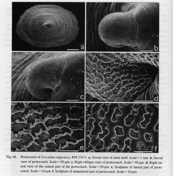
Cocculina nipponica Kuroda and Habe, 1949
(Figs. 68 a-f, 69 a-d, 70 a-b, 71, 72 a-f)
Protoconch: Protoconch planispiral, symmetrical in dorsal view (Figs. 68 a-c). Surface ornamented with network pattern, coarser laterally, denser dorsally (Figs. 68 d-f). Protoconch-teleoconch boundary marked by very thin, slightly undulated apertural lip. Growth direction of protoconch parallel to anterio-posterior axis of teleoconch.
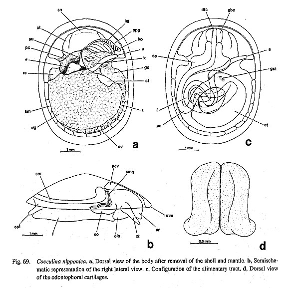
External Anatomy: Animal limpet-shaped. Mantle margin lacking circumpallial microtentacles. Shell muscle (sm) horseshoe-shaped, penetrated by afferent mantle vessels (Fig. 69 a), thickened at anterior ends by fusion with head retractor muscles, forming somewhat incurved rounded projections.
Head bearing short snout (sn) and non-papillate cephalic tentacles (ct). Mouth surrounded by bristles. Outer lip with broad oral lappets (ola) laterally. Eyes absent. Copulatory organ on right neck region (co. Fig. 69 b).
Paired, ciliated, non-papillate epipodial tentacles (ept) present on posterior part ofepipodium (Fig. 69 b). Epipodial sense organs absent. Operculum absent.
Pallial Complex: Pallial cavity shallow, occupying less than half of body length, containing pseudoplicate gill on left side, left osphradium, single kidney opening, anus, and left hypobranchial gland. Most of area of posterior mantle roof occupied by pericardium on left and kidney on right. Folded, pseudoplicate gill (ppg) arising on right mantle roof (Fig. 69 a), covered by somewhat irregular ciliation, Hypobranchial gland (hg) on left side of gill.
Digestive System: Odontophore fixed by lateral and ventral protractors (Figs. 70 a, b): (1) Outer part of lateral protractors (lp) running from posterior ends of cartilages to wall of snout. Inner part of muscle connecting with ventral extensions of cartilages and buccal constrictor covering oral tube. (2) Outer ventral protractors (ovp) originating from body musculature below buccal mass, inserting on posterior extensions of cartilages. Inner ventral protractors (ivp) on dorsal side of outer protractors, similar in configuration to
outer ventral protractors. Left and right halves of odontophore united by postdorsal buccal tensor (pdt) and dorsal buccal tensors (dbt). Levators and depressors of odontophore absent.
Paired jaw plates (j) distinguished by darker color from adjacent area of oral tube, with smooth anterior margin and inner surface lacking scale-like sculpture.
Radular sac (rds) bent ventrally behind buccal mass (Fig. 71). Posterior end bifurcated to form globular lateral projections (Figs. 70 a, b). Radular formula n-(1+3)-0-(3+1)-n. Each transverse row symmetrical. Central area of radula lacking teeth, with broad exposed space on radular membrane marked by longitudinal ridges, corresponding to radular segments. Inner lateral teeth narrowly elongated. Second laterals with shorter shaft and more extended base than innermost laterals; cusp tri-denticulate with innermost denticle longest. Third laterals elongated, sickle-like; cusp with single acute hook. Outer (fourth) laterals elongated, stout; dorsalsurface of cusp streaked by fine longitudinal lirae converging distally; inner basal corner articulating with base of third lateral teeth. Lateromarginal plates absent. Marginal teeth broad basally, tapering in distal third; cusps sickle-shaped, denticulate, with sharply elongated denticles.

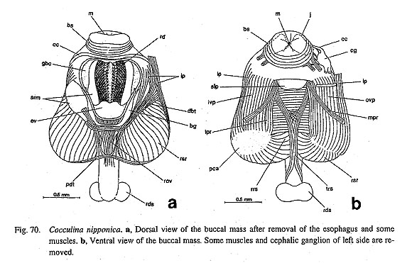
Action of radula controlled by two pairs of protractors and single pair of retractors of subradular membrane. Due to development of odontophoral cartilages and retractor muscles, buccal mass markedly swelling in posterior part. Median protractors (mpr) emerging from ventral side of subradular pouch, attaching to ventral body musculature. Lateral protractors (lpr) originating from posterior extensions of cartilages.
Buccal mass containing pair of odontophoral cartilages (ca) (Figs. 69 d, 71,72 a, b) connected by single-layered ventral approximator muscle. Tensor of cartilages absent.

Ventral side of oral tube forming simple sublingual pouch (slp) without lateral glandular outgrowths (Fig. 70 b). Buccal cavity extending laterally, its wall lined by single layer of tall glandular cells. Radular diverticulum narrow. Salivary glands absent.
Anterior esophagus depressed over odontophore. Interior clearly divided into dorsal food channel (dfc) and lateral pouches by dorsal and ventral folds. Behind buccal mass, lateral pouches developing into large, flat pouches (ep). Both dorsal and ventral folds disappearing in posterior esophagus (pe). Inner wall of mid
esophagus smooth. Esophagus gradually curving left, recurving anterodorsally toward stomach.
Stomach (st) inverted C-shaped (Fig. 69 c). Part of stomach appearing on dorsal surface of visceral hump, mostly embedded within digestive glands (dg). Digestive glands opening into stomach through single pore at initial dorsal part. Inside of stomach ventrally curved by typhlosoles and corrugated sorting area. Dorsolateral wall entirely smooth with cuticularized gastric shield. Tooth of gastric shield (gst) projecting on dorsal part just behind opening of digestive glands. Intestine (i) bending four times at more ventral level than posterior esophagus and stomach; only terminal part running along dorsal surface, Recum not penetrating pericardium or ventricle. Anus (a) exceeding right anterior limb of shell muscle.
Circulatory System: Pericardium (pc) on left side just posterior to pallial cavity. Heart consisting of single auricle and single ventricle (Fig. 69 a). Auricle (au) lying on right anterior side, connecting with ventricle through narrow opening. Auricle receiving blood from pseudoplicate gill. Connection with kidney uncertain. Ventricle (v) rather freely fixed within pericardium on left posterior side of auricle. No clear boundary identified between anterior and posterior aortae.
Excretory System: Single kidney (k) on right side of pericardium. Anterior part of kidney mainly attached to mantle skirt (Fig. 69 a). Posterior part overrun by rectum. Small kidney opening (ko) to left side
of anus beneath hypobranchial gland (Fig. 72 c). Internal microstructure not observed histologically.
Reproductive System: Hermaphroditic gonad on ventral side of visceral mass (Fig. 71). Testis (t) and ovary (ov) partly separated, but intermingled with each other on ventral side (Figs. 71,72 f). Gonoduct (gd) originating from right side of gonad, extending along shell muscle. Distal part of gonoduct dilated, highly glandular (Fig. 72 d). Near genital opening, another duct extending toward left side under pericardium. Terminal part of duct enlarged to form receptaculum seminis (rs), partly appearing on left dorsal surface between pericardium and shell muscle (Figs. 69 a, 72 e). Near genital opening, deep seminal groove (smg) running along edge of shell muscle, extending to verge of copulatory organ (co) continuously. Copulatory organ arising behind base of right cephalic tentacle.
Nervous System: Circumesophageal nerve ring hypoathroid. Cerebral ganglia at bases of cephalic tentacles, connected by cerebral commissure. Pleural and pedal ganglia (pdg) under buccal mass (Fig. 71), united with cerebral ganglia (cg) by separate, short cerebropleural and cerebropedal connectives.
Cerebral ganglia provided with at least four branches to oral region, but lacking labial commissure and ganglia (Fig. 70 b). Cerebrobuccal connectives arising from inner ventral sides of cerebral ganglia, running along anterolateral edges of cartilages. Buccal ganglia (bg) visible over retractors of subradular membrane (rsr) when esophagus removed dorsally (Fig. 70 a), mainly innervating esophagus anterodorsally and buccal musculature both ventrolaterally and posteriorly.
Structural details of visceral nerve loop and innervation to pallial organs not observed. Pedal ganglia extending thick pedal cords, apparently lacking commissures. Statocysts attached to inner anterodorsal sides of pedal ganglia.
Family Neritidae Rafinesque, 1815
Nerita (Theliostyla) albicilla Linnaeus, 1758
(Figs. 73 a-b, 74 a-d, 75 a-e, 76 a-d, 77 a-h,78 a-b)
Protoconch: Protoconch globular, multispiral, convoluted in orthostrophic direction (Figs. 78 a, b). Outer surface free of prismatic deposits, completely smooth except for fine growth lines. Protoconch-teleoconch boundary clearly demarcated. Margin of protoconch aperture slightly thickened.

External Anatomy: Head-foot nearly symmetrical. Visceral hump not spirally coiled, with apex directed toward anterior. Mantle margin smooth, without microtentacles.
Head with short snout (sn), non-papillate cephalic tentacles (ct), eyestalks (es), and right cephalic penis (pen) in male. Outer lip of mouth smooth, laterally extended to form oral lappets. Eyestalks with lateral extension arising from outside of cephalic tentacles. Closed eyes on tips of eyestalks. Flap-like penis to inside of right cephalic tentacle.
Epipodial region of foot lacking epipodial tentacles and sense organs. Attachment of shell muscle divided into right and left portions (lsm, rsm), each subdivided into nine or ten bundles (Figs. 73 a, b). Right muscle attaching to inside of apical area; left muscle to basal side of columella.
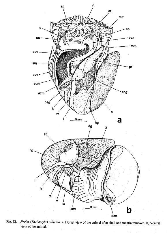
Pallial Complex: Pallial cavity very deep, reaching almost entire body length, containing left ctenidium, right "vestigial gill," left osphradium, left kidney opening, anus, genital opening(s), and right hypobranchial gland.
Single (left) bipectinate ctenidium arising from left posterior pallial wall. Ctenidium supported by short afferent and efferent ctenidial membranes. Afferent and efferent ctenidial axes containing afferent and efferent ctenidial vessels, ctenidial nerves, and retractor muscles (Fig. 77 a). Ctenidial lamellae triangular-shaped in alternating arrangement. Midline of lamellae projecting vertically, generating ridged appearance. Efferent side of ctenidial lamellae lacking skeletal rods (Fig. 77 b). Surface of ctenidial lamellae covered with zoned series of frontal, abfrontal, and lateral cilia. Bursicles absent.
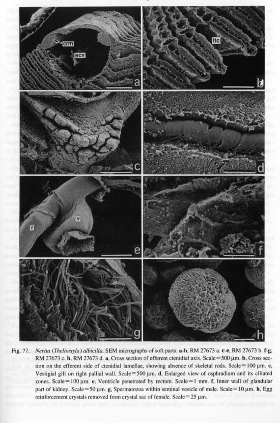
Marked tuberculation of "vestigial gill" (Fretter, 1965) on right side of kidney opening (Fig. 77 c). Cili
ary bands on surface of gill absent. Gill internally connected with large space of efferent pallial vessel.
Osphradium (os) resting on mantle skirt on anterior side of attachment of efferent ctenidial axis (Fig. 73 a), with long vermiform ridge accompanied by furrow and ciliated band on each side (Fig. 77 d). Hypobranchial gland (hg) well-developed on right side, associated with genital duct.
Digestive System: Oral tube surrounded by buccal sphincter and constrictor (bs, bct), mandibular protractors and retractors (mp, mr), and buccal tensor muscles (bt).
Buccal mass anchored on musculature of body wall by lateral protractors (lp), ventral protractors (vp), median and dorsal levators (ml, dl), posterior depressors (pd), anterior levators (al), and anterior tensors (at) (Figs. 74 a-d, 75 a). Posterior part of odontophore united by postdorsal buccal tensor (pdt). Posterior levator and dorsal buccal tensor absent.
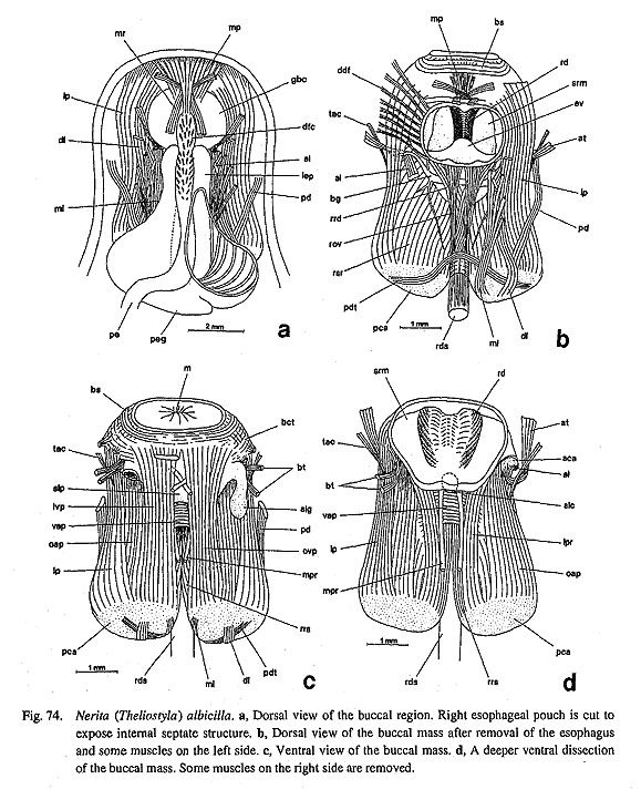
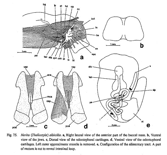
Jaws (j) completely adhered to dorsal area of oral tube, composed of distinctly paired translucent plates fused by median cuticularized zone (Fig. 75 b). Each plate subrectangular with rounded edge. Anterior margin and inner surface lacking sculpture and projection.
Radular sac very long, coiling at least three times. Posterior end of radular sac not clearly divided. Radular formula n-(1+3)-1-(3+1)-n. Radular row symmetrical. Central tooth laterally rectangular. First lateral teeth with inner margin completely fitting into central tooth; cusp small, reflected in flap-like, triangular form at outer anterior corner. Second and third laterals in narrow groove between first inner lateral and outer lateral teeth. Outer (fourth) laterals greatly enlarged transversely; cusp thickened, smooth, shield-shaped; base inflated with deep pit on outer side. Lateromarginal plates absent. Marginals with long shafts and smooth cusps.
Subradular membrane attached anteriorly with two pairs of protractors, median and lateral protractors (mpr, lpr), and laterally with well-developed retractor muscles (rsr). Retractors of radular sac (rrs) originat
ing from inner sides of posterior cartilages, inserting on ventral plane of radular sac. Postmedian retractors and tensor of radular sac absent.
Odontophoral cartilage consisting of anterior, posterior, and median pairs (Figs. 75 c, d). Anterior cartilages (aca) elongated, attached posteriorly to posterior cartilages (pca). Unpaired median cartilage (mca) lying on median grooves between paired anterior cartilages. Small sublingual cartilage (slc) below anterior tip of subradular membrane (Fig. 74 d). Ventral sides of anterior cartilages connected to ventral approximator (vap). Anterior and posterior cartilages longitudinally linked with outer approximators (oap). Sides of anterior cartilages attached to body wall by thin tensor muscles (tac) (Figs. 74 b-d, 75 a).
Licker absent on anterior tip of subradular membrane (Fig. 74 d). Inside of oral tube thickened by cuticu-larized epithelium. Sides of buccal pouches especially thickened by glandular epithelium composed of tall columnar cells. Salivary glands not differentiated around buccal cavity. Paired sublingual glands (slg) pro-jecting on sides of sublingual pouch (Fig. 74 c). Radular diverticulum deeply formed.
Anterior esophagus dorsoventrally depressed with paired lateral esophageal pouches (lep) and median dorsal food channel (dfc). Lateral esophageal pouches gradually increasing in width posteriorly, forming large spaces; inner wall covered with separate posterior esophageal glands (peg) (Fig. 74 a). Posterior part of pouches truncated from main trunk of esophagus. Remaining part of esophagus reducing in diameter, forming posterior esophagus (pe) with inner surface sculptured by longitudinal furrows.
Stomach (st) well-inflated, oval (Fig. 75 e), with right side exposed on surface of visceral hump in ventral view. Corrugated pattern of sorting area partly visible externally as fine lines (Fig. 73 b). Gastric caecum small, crescent-shaped. Interior of stomach containing paired openings of digestive glands, large cu-ticularized gastric shield, tooth of gastric shield, ciliated sorting area, and intestinal groove between major and minor typhlosoles. Opening to intestine lying on left posterior side of stomach.
Intestine (i) with complicated course, forming anterior loops twice over posterior esophagus (Fig. 75 e). Rectum penetrating pericardium and ventricle (Fig. 77 e), running along genital ducts, ending as anus (a) on anteroventral side of genital pore.
Circulatory System: Posterior end of pericardium deeply elongated into visceral region along left shell muscle. Heart consisting of paired unequal auricles and median ventricle (Fig. 73 b). Right auricle (ra) re-duced but functional, connected to renal wall. Left auricle (la) well-developed, receiving blood from ctenidium and mantle skirt. Inner wall of ventricle firmly supported by muscular strands. Blood vessel from ventricle bifurcating into anterior and posterior aortae. Bulbous aorta not formed.
Excretory System: Single kidney (k) on posterior side of ctenidium (Fig. 73 a). Internal wall distinctly partitioned into two portions. Glandular part on right side filled with folded walls (Fig. 77 f), extending gradually toward anterior right corner; left side of non-glandular area forming larger smooth space. Renop-ericardial duct running toward right side of kidney; renopericardial pore perforating anterior section of glandular zone of kidney. Opening of kidney represented by vertical slit.
Reproductive System: Gonad lying on dorsal end of visceral hump (Figs. 73 a, b). Sexes separate. In both testis and ovary, gametes aggregating into cylindrical clusters of similar form; ovary distinguished by black epithelium in contrast to non-pigmented epithelium of testis. Gonoduct not connected to kidney. Pallial gonoduct showing remarkable sexual dimorphism.
Male Gonoduct (Figs. 76 a, b): Vas deferens (vd) from testis totally covered with dark gray epithelium, distal part especially forming very complicated entangled mass of ducts below hypobranchial glands, serving as seminal vesicle (sv). Extended part of seminal vesicle including completed spermatozoa (Fig. 77 g) packed within spermatophore, exhibiting whitish hue. Course of seminal vesicle completely random; terminal part of vas deferens invariably opening into pallial gonoduct from ventral side. Most of pallial gonoduct covered ventrally by prostate gland (pr), divided by ridges internally. Middle part of prostate twisted to
form crescent-shaped prostate pouch. Dorsal side of prostate attached by so-called basal gland (bsg) com-posed of translucent gelatinous tissue of large glandular cells. Posterior half of prostate covered by so-called annex gland (ang) both ventrally and dorsally. Ventral lobe of annex gland covering larger area of prostate than dorsal side. Ciliated sperm groove between gonopore and cephalic penis absent.

Female Gonoduct (Figs. 76 c, d): Female gonoduct with two genital openings (diaulic), consisting of three major parts: (i) spermatophore sac (sps) and receptaculum seminis (rs) for reception and storage of sperm; (ii) oviduct (ovd) as conduit of egg cells; (iii) pallial oviduct for production of egg capsule enclosing fertilized eggs.
Spermatophore sac opening as vagina (va). After copulation, spermatophores conveyed into spermato-phore sac with rounded part directed posteriorly. Long filamentous tip penetrating far back into chamber of receptaculum seminis. Ducts of receptaculum seminis (drs) composed of three parts: (i) Proximal part arising anteriorly from spermatophore sac, posteriorly followed by receptaculum seminis. (ii) Middle part be-coming extremely thin compared to other parts, (iii) Distal part recurved, becoming thick again, leading to ventral albumen gland (vag), inflating in middle to form right-directed extension pigmented black internally (Fig. 76 d), of unknown function.
Posterior end of pallial gonoducts comprised of albumen glands divided into two parts. Ventral albumen gland (vag) mainly lying on ventral side, receiving sperm from ducts of receptaculum seminis. Dorsal albumen gland (dag) partly overlying ventral gland, connected to oviduct from ovary. Boundary between ventral and dorsal glands with particular glandular zone staining darkly in methylene blue. More anteriorly, pallial oviduct mainly consisting of capsule gland (cpg). Crystal-secreting sac (=crystal sac) (cs), attached to termination of gonoducts, connected to gonoduct only near female opening anteriorly. Sac distended in breeding season by egg reinforcement crystals (Fig. 77 h), becoming clearly visible from dorsal surface by whitish color. Female opening (fo) simple pore; opening of vagina slit-like.
Nervous System: Circumesophageal nerve ring hypoathroid. Cerebral ganglia (cg) situated at bases of cephalic tentacles. Pleural and pedal ganglia fused, forming massive complex. Pedal ganglia closely lo-cated; pedal commissure represented by short and broad contact zone. Right and left pleural ganglia con-nected by thick pleural commissure.
In buccal region, cerebral ganglia connected to cerebral commissure (cc) in front of buccal cavity (Fig. 75 a). Cerebral ganglia giving off branches of oral nerve (on) anteriorly, and tentacular and optic nerves (tn, opn) laterally. Nerve to cephalic penis arising from beneath sublingual pouch; labial commissure (Ic) con-necting right and left cerebral ganglia. Labial ganglia absent. Cerebrobuccal connectives branching from labial commissure, not directly from ventral extension of cerebral ganglia. Buccal ganglia (bg) connected with buccal commissure over odontophore between anterior esophagus and radular diverticulum.
Visceral loop forming very loose loop in accordance with deep pallial cavity. Origin of loop only from right pleural ganglion. Subesophageal part of loop much thicker than supraesophageal part, the latter arising from more anterior part of right pleural ganglion than the former. Below pallial gonoduct and "vestigial gill," subesophageal ganglion supplying nerves to genital duct, gonad, and right shell muscle. Loop further continuing to visceral ganglion lying below kidney opening, innervating kidney, pericardium, and digestive tract.
Very thin supraesophageal loop ascending to right side of buccal mass, passing over esophagus, finally reaching base of ctenidium where it diverges into anterior and posterior parts. Anteriorly directed part en-tering base of efferent ctenidial membrane, also innervating ctenidium and osphradium. Posteriorly directed part representing viscero-supraesophageal connective, running along floor of pallial cavity, uniting with visceral ganglion.
Lateral body walls receiving dense innervation directly from pleural ganglia. Thick nerves especially penetrating anterior rims of shell muscles, extending mantle margin circularly, forming zeugoneury with supraesophageal loop on left side.
Pedal ganglia with thick pedal cords with many fine pedal nerves to pedal musculature. From anterior part of pedal ganglia, pair of thin anterior pedal nerves extending toward pedal region below head. Stato-cysts on dorsal sides of pedal ganglia.
Nerita (Puperita) bensoni (Récluz, 1850)
(Figs. 78 c-d)
Protoconch: Protoconch globular, multispiral, orthostrophic. Surface completely smooth except for fine growth lines. Protoconch terminating in clear line, followed by teleoconch sculptured by axial growth lines and about 40 spiral cords.
Nerita sp.
(Figs. 78 e-f)
Protoconch: Protoconch globular, multispiral, with fine growth lines. Coiling direction orthostrophic. Early teleoconch sculptured by about 35 thick, rough, granulose radial riblets.
Pisulina adamsiana G. and H. Nevill, 1869
(Figs. 78 g-h)
Protoconch: Protoconch globular, multispiral, orthostrophic. Fine growth lines visible on exterior sur-face, details unclear in worn empty shells examined. Protoconch mostly covered by teleoconch, with only small apical area exposed.
Septalia porcellana (Linnaeus, 1758)
(Figs. 79 a-d, 80 a-b)
External Anatomy: Animal limpet-shaped. Mantle margin simple, without microtentacles. Attachment area of shell muscle horseshoe-shaped, interrupted posteriorly (Fig. 79 a). Muscle not divided into bundles or penetrated by blood vessels.
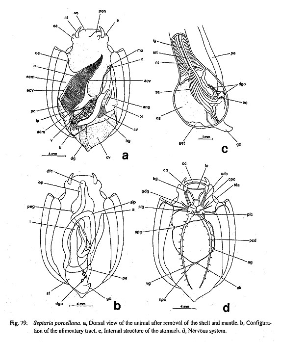
Head with short snout (sn), paired non-papillate cephalic tentacles (ct), eyestalks (es), and right cephalic penis (pen) in male. Outer lip enlarged into broad disk with extended oral lappets. Eyestalks laterally ex-tended, broader than cephalic tentacles. Eyes (e) closed with cornea. Cephalic penis arising from right side; anterior ventral margin marked by deep groove.
Epipodium lacking sensory projections of epipodial tentacles and sense organs. Calcified operculum in space between foot and visceral hump, buried in pedal musculature anteriorly.
Pallial Complex: Pallial cavity very deep, reaching 80% of body length in deepest section, containing left ctenidium, left osphradium, left kidney opening, anus, genital opening(s), and right hypobranchial gland (Fig. 79 a). "Vestigial gill" absent on right side (unlike in Nerita).
Ctenidium (c) bipectinate. Both afferent and efferent axes widely supported by broad afferent and efferent membranes. Ctenidial lamellae depressed, with apices revealing weak ridges medially. Efferent side of lamellae lacking skeletal rods and bursicles.
Osphradium (os) with lateral ciliated zones lying along anterior end on right side of shell muscle in ex-tension line of efferent ctenidial membrane. Hypobranchial gland (hg) associated with genital duct on right side.
Digestive System: Buccal musculature identical in composition with that in Nerita. Jaws composed of paired plates on dorsal side of oral tube, connected medially to filmy inner surface. Surface and anterior margin smooth.
Radular sac long, with several loops; posterior end simple. Radular formula n-(1+3)-1-(3+1)-n.
Radular row symmetrical. Central tooth rectangular with shortly stalked base; non-denticulate cusp clearly formed by thick reflection. First inner laterals transversely elongated; posterior margin of base concave, with backward extension near outer corner; cusp triangular with rounded tip. Second and third laterals ex-tremely reduced relative to other teeth, longitudinally narrow with indistinct cusps. Outer (fourth) laterals largest in radular row; cusp laterally thickened in shield-like shape; cutting edge lacking denticles; shaft stout, robust, with deep basal hole; cusp continuing to membranous extension of shaft whose margin sig-moidally bent. Lateromarginal plates absent. Marginal teeth with elongated shafts and finely denticulate cusps.
Odontophore containing paired anterior, posterior, and median odontophoral cartilages. Sublingual carti-lage absent (unlike in Nerita).
Licker absent. Sublingual pouch well-developed, with sublingual glands projecting on outer sides. Radu-lar diverticulum clearly present. Salivary glands not differentiated around buccal cavity. Anterior esophagus depressed over odontophore. Lateral pouches (lep) followed by enlarged and septated posterior esopha-geal glands (peg) (Fig. 79 b). Mid-esophagus not clearly distinguished. Posterior esophagus (pe) very long, extending almost straight toward stomach at end of visceral region. Paired digestive glands (dg) with couple of ducts from stomach. Posterior wall of stomach giving rise to short gastric caecum (gc) (Fig. 79 c). Tooth of gastric shield (gst) lying on dorsal side. Sorting area (sa) and intestinal groove (ig) between major and minor typhlosoles (mt, nt) well defined on ventral side. Intestine (i) forming double loops anteriorly (alp), penetrating pericardium and ventricle, finally ending as anus (a) on left side of gonoduct.
Circulatory System: Pericardium lying in triangular area between ctenidium and kidney (Fig. 79 a). Pos-terior part of pericardium extending below visceral hump as tubular sac. Heart, consisting of greatly asym-metrical pair of auricles and median ventricle. Left auricle (la) well-developed, receiving blood fromctenidium. Right auricle greatly reduced to thin string-like form.
Excretory System: Kidney (k) mostly occupying posterior part of mantle roof, extending into visceral re-gion along rectum (Fig. 79 a). Lumen divided into lamellate glandular area on right side and smooth non-glandular area on left side. Renopericardial duct passing from anterior right corner of pericardium into ante
rior chamber of glandular part of kidney. Kidney opening short slit.
Reproductive System: Gonad lying over digestive glands mainly on right side. Gonoduct not connected with kidney, showing extreme sexual dimorphism.
Male Gonoduct (Fig. 80 a): Vas deferens (vd) folded complexly as mass of innumerable loops in posterior end of pallial gonoduct. Proximal loops distended with sperm, where vas deferens functioning as seminal vesicle (sv). Most of pallial gonoduct occupied by prostate gland (pr). Seminal vesicle leading ventrally into prostate; their connection invisible externally due to covering of annex gland (ang). Basal gland (as in Nerita) absent. Prostate curving twice before opening in anterior pallial cavity. Ciliated sperm groove between gonopore to cephalic penis absent.
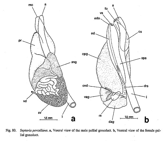
Female Gonoduct (Fig. 80 b): Female gonoduct with three genital openings (triaulic). Vagina (va) opening at tip of spermatophore sac (sps). From posterior lateral wall, duct arising through elongated slit, bifur-cating at base into enigmatic duct (ed) anteriorly and duct of receptaculum seminis (drs) posteriorly. Enigmatic duct running anteriorly along spermatophore sac, discharging into pallial cavity at minute pore (edo). Duct of receptaculum roughly nodose on external surface, posteriorly branching into receptaculum seminis (rs) and duct to ventral albumen gland. Dorsal and ventral albumen glands (dag, vag) divided by median zone staining darkly in methylene blue. Most of pallial oviduct occupied by capsule gland (cpg). Crystal sac (cs) communicating with oviduct at distal end. Opening of gonoduct (fo) lying beside anus.
Nervous System: Circumesophageal nerve ring hypoathroid (Fig. 79 c). Cerebral ganglia (cg) at bases of cephalic tentacles. Cerebral commissure (cc) in front of buccal cavity. Pedal and pleural ganglia (pdg, plg) united as large paired mass behind buccal mass. Pedal ganglia connected by thick commissure. Pleural gan-glia dorsally united by distinct pleural commissure (plc) whose right origin lies more posteriorly than that of left side.
In buccal region, labial ganglia absent, but distinct labial commissure (lc) connecting to cerebral ganglia ventrally behind mouth. Buccal ganglia (bg) arising from labial commissure, not directly from cerebral ganglia. Nerve to cephalic penis arising from inner part of right cephalic ganglion.
Visceral loop arising only from right pleural ganglion. Supraesophageal part of visceral loop much thinner than subesophageal part The former running over buccal mass, while the latter extending along lateral body wall. Visceral ganglion (vg) lying on right side of kidney opening. Extremely thin viscero-supraesophageal connective forming complete visceral loop.
Pedal cords (pcd) giving off few branches. Pedal commissure indistinct. Statocysts (sta) attached to dorsal sides of pedal ganglia below pleural commissure.
|