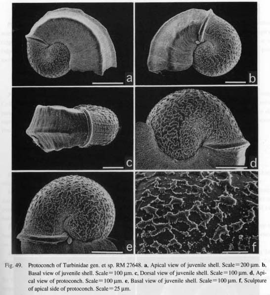
Turbinidae gen. et sp.
(Figs. 49 a-f)
Protoconch: Protoconch paucispiral, planispiral. Lateral pouch not clearly formed. Suture of protoconch short, clear. Sculpture consisting of discontinuous spiral thread and zigzag ridge-like deposits both of which partly anastomose to form reticulate pattern; interspace powdered with minute grains. Sculpture of central area finer than that of periphery both on apical and basal sides. Proloconch-teleoconch boundary clearly demarcated by prominent axial ridge.
Homalopoma nocturnum (Gould, 1861)
(Figs. 50 a-d)
Protoconch: Protoconch paucispiral, almost planispiral, slightly hyperstrophic. Lateral pouch not in-flated; constriction of suture producing shallow depression. Outer surface ornamented with fine irregular microtubercles that aggregate to construct axial sculpture. Fine striae also formed in radial direction on both apical and basal sides. Protoconch-teleoconch boundary marked by constriction.
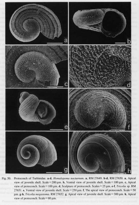
Tricolia sp.
(Figs. 50 e-f)
Protoconch: Protoconch oblong, conispirally coiled. Extension of lateral pouch indistinct. Suture marked by shallow constriction occurring inside apertural area. Protoconch sculptured by clear spiral ridges; number initially five, increasing with growth, attaining ten at protoconch aperture. Interspace of ridges forming reticulation with ridge-like deposits and round pit-like depressions. Apertural margin of pro-toconch weakly thickened, clearly distinguished from spiral cords of early teleoconch.
Tricolia megastoma (Pilsbry, 1895)
(Figs. 50 g-h)
Protoconch: Protoconch discoidal, paucispiral, well-inflated. Lateral pouch on apical side not prominent. Coiling direction orthostrophic in both protoconch and teleoconch. Sculpture showing very characteristic pattern of three distinct spiral ridges in peripheral margin. Each ridge gradually bifurcating, obliquely directing toward outer periphery. Interspace between two ridges totally smooth even at high magnification. Protoconch-teleoconch boundary delimited by simple axial line.
Family Trochidae Rafinesque, 1815
Chlorostoma lischkei (Tapparone-Canefri, 1874)
(Figs. 51 a-b, 52 a-b, 53 a-e)
External Anatomy: Visceral hump spirally coiled in 2.5 volutions. Mantle margin (mm) simple, lacking microtentacles. Columellar muscle (cm) longitudinally elongated, narrowly restricted to columellar side.
Head with short snout, cephalic lappets, cephalic tentacles, and eyestaiks. Outer lip of mouth thickened, forming circular oral disk. Cephalic lappets transversely elongated, divided into distinct pairs. Eyeslalks on posterior sides of cephalic tentacles. Eyes opening externally. Neck lobes present on inhalant (left) and ex-halant (right) sides.
Epipodium provided with four pairs of very long epipodial tentacles (ept) extensible over periphery of body whorl. Surface of tentacles papillate with ciliary crown. Base of each tentacle with knobby projection on which epipodial sense organ lies. Multispiral corneous operculum on epipodium.
Pallial Complex: Pallial cavity reaching more than one-half volution of body whorl, containing left ctenidium with osphradium, paired kidney openings, anus, and paired hypobranchial glands (Figs. 51 a, b).
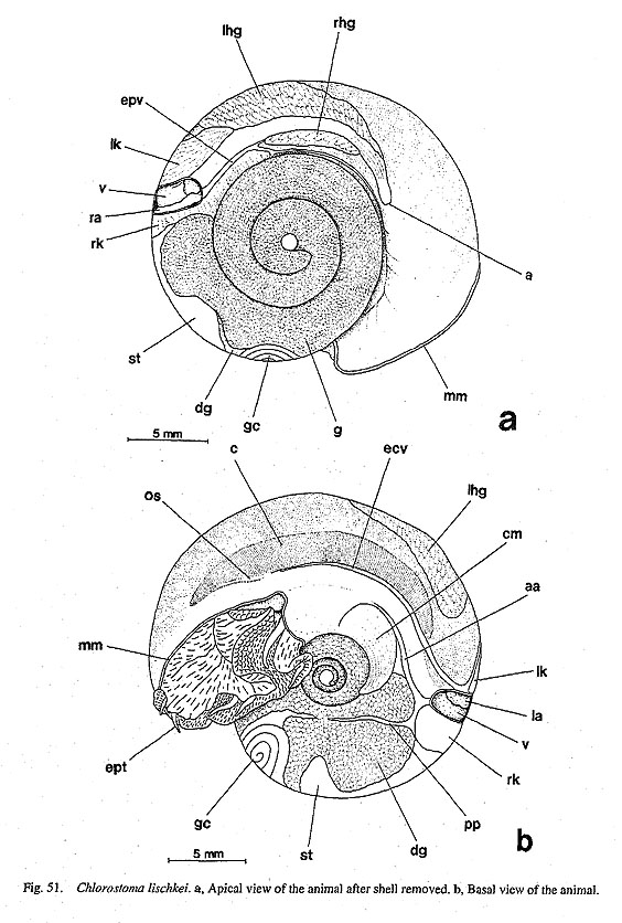
Ctenidium (c) bipectinate, long, extending along left wall of pallial cavity. Efferent (left) side supported by long efferent ctenidial membrane except anterior free portion; afferent side suspended by afferent mem-brane, attached to mantle skirt along left margin of hypobranchial glands.
Afferent and efferent axes include afferent and efferent ctenidial vessels, also paired retractor muscles and median nerve. Efferent side of anterior free portion stiffened by skeletal rods that are distinguished by reddish staining in histological sections.
Ctenidial lamellae depressed in form like acute-angled triangle; apex of lamellae directed and projected to afferent side. Surface with frontal, abfrontal, and lateral cilia. Efferent side of lamellae supported by paired skeletal rods. Bursicles prominent on efferent side of lamellae. Osphradium on outer surface of anterior free portion of efferent axis.
Hypobranchial glands paired, extremely unequal in size. Right gland (rhg) elongated along rectum. Left gland (lhg) extending in posterior median direction, contacting anterior part of left kidney. Transverse pallial vein running below left gland.
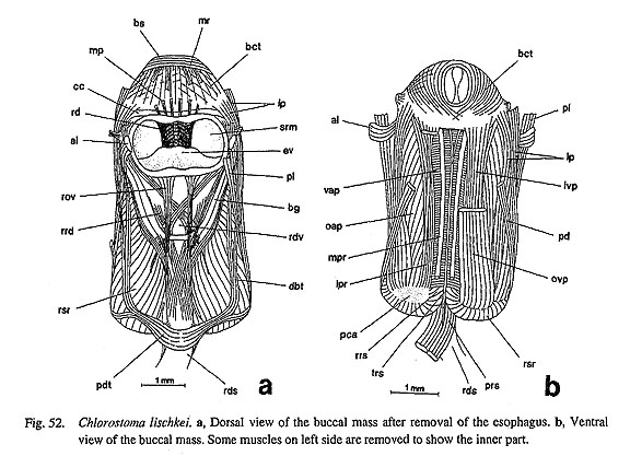
Digestive System: Oral tube surrounded by buccal sphincter (bs) and constrictor (bct) with mandibular protractors (mp) and retractors (mr). Odontophore fixed by lateral protractors (lp), outer and inner ventral protractors (ovp, ivp), anterior levators (al), posterior levators (pl), and posterior depressors (pd) (Figs. 52 a,
b). Posterior side of odontophore bound by postdorsal buccal (pdt) and dorsal buccal tensors (dbt).
Jaws paired, subtrapezoid, thin, nearly entirely attached to wall of oral tube. Anterior margin and inner surface totally smooth (Fig. 53 c).
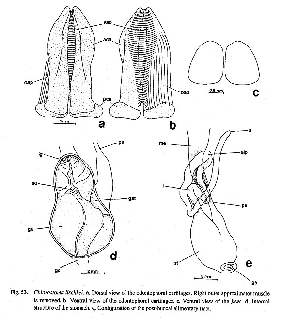
Radular sac forming loops with few volutions on right ventral side of mid-esophagus. Posterior end clearly bifurcated. Radular formula n-5-1-5-n. Tooth row symmetrical. Central tooth subrectangular, plate-like, with lateral extensions and slightly reflected cusp. Lateral teeth with keeled shafts and acute cusps; bases with prominent outward projection and intimate articulation with each other. Protolateromarginal plates smoothly extended from bases of innermost marginals. Marginal teeth all very long; cusps with weak median ridge and serration on outer side.
Subradular membrane attached by median and lateral protractors (mpr, lpr) on anterior side and by retractors (rsr) on lateral and posterior sides (Fig. 52 b). Radular sac ventrally inserted by retractors (rrs) and postmedian retractors (prs). Tensor of radular sac (trs) running from dorsal side of sublingual pouch to ventral side of radular sac between paired postmedian retractors (prs).
Odontophore containing two pairs of cartilages (Figs. 53 a, b). Anterior cartilages (aca) longitudinally elongated, posterior cartilages (pca) transversely extended. Ventral sides of anterior cartilages connected by single-layered ventral approximator along length. Anterior and posterior pairs united by outer approxima-tors (oap).
Sublingual pouch shallow, lacking glandular projection. Licker smooth on anterior end of subradular membrane. Radular diverticulum present. Salivary glands without well-developed ducts over dorsal surface of buccal cavity, partly overlapping dorsal food channel.
Dorsal food channel well-inflated, merging into mid-esophagus. Initial part of mid-esophagus (me) enlarged by esophageal pouches. Inside of mid-esophagus entirely covered by papillate glandular epithelium
except in ciliated zone between paired dorsal and ventral folds. Posterior esophagus (pe) formed by con-striction of mid-esophagus; inner surface longitudinally corrugate.
Stomach (st) large, well-expanded (Fig. 53 e), strongly bent in V-shape, divided into right proximal and left distal parts. Both ventral and dorsal surfaces of stomach covered by digestive glands (dg); lateral wall of distal parts widely exposed on surface of visceral hump (Figs. 51 a, b). Posterior apex giving rise to spiral gastric caecum (gc) consisting of three volutions. Inside of proximal part mainly occupied by smoothly cuticularized gastric shield (gs) (Fig. 53 d). Tooth of gastric shield (gst) on dorsal side of openings of esophagus and digestive glands. Sorting area (sa) weakly developed in median area. Typhlosoles and intes-tinal groove (ig) well-defined in distal end of stomach.
Intestine (i) forming single anterior loop on right side (Fig. 53 e). Rectum penetrating pericardium and ventricle, running on roof of pallial cavity between paired kidneys and hypobranchial glands.
Circulatory System: Pericardium lying transversely along posterior wall of pallial cavity, containing paired asymmetrical auricles and single ventricle (Figs. 51 a, b). Right auricle (ra) thinly elongated on right dorsal side. Left auricle (la) on left ventral side larger than right one. Right auricle receiving blood only from efferent pallial vein; left auricle connecting efferent ctenidial vessel and efferent renal vein.
Excretory System: Excretory system consisting of paired kidneys of different form and histology (Figs. 51 a-b). Right kidney (rk) lying posterior to pericardium on dorsal surface, also extending internally toward anterior right visceral region. Right kidney opening projecting as short papilla. Left kidney (lk) entirely lying in pallial cavity, internally exhibiting condition of so-called papillary sac; left kidney opening repre-sented by longitudinal short slit. Renopericardial ducts paired.
Reproductive System: Gonochoristic gonad (g) lying over digestive glands, extending over stomach and part of right kidney when ripe (Fig. 51 a). Gonoduct running inner columellar side, opening into right kidney. Gametes discharged into pallial cavity through right excretory pore.
Nervous System: Circumesophageal nerve ring hypoathroid. Labial region innervated by several nerves from cerebral ganglia, lacking distinct labial commissure and ganglion. Visceral loop arising from right and left pleural ganglia. Left nerve from supraesophageal to osphradioctenidial ganglia connected by nerve from left pleural ganglion (zeugoneury). Pedal cords scaraliform. Statocysts on anterior part of pedal ganglia.
Chlorostoma. sp.
(Figs. 54 a-c)
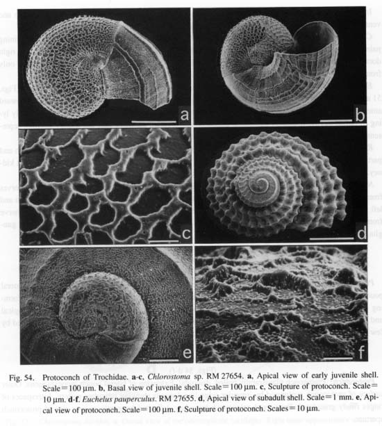
Protoconch: Protoconch coiled in slightly hyperstrophic direction with about 1.25 volutions. Lateral pouch insignificant on apical side, with unclear suture. Basal side weakly inflated, with lateral pouch forming ventral extension. Protoconch surface totally covered with reticulate sculpture that is denser in apical and basal regions than at periphery. Protoconch-teleoconch boundary marked by thin axial rib followed by few spiral cords of early teleoconch.
Euchelus pauperculus (Lischke, 1872)
(Figs. 54 d-f)
Protoconch: Protoconch planispirally coiled, discoidal. Extension of lateral pouch inconspicuous. Outer surface of protoconch sculptured by irregular ridge-like deposits showing reticulate pattern. Interspaces of ridges finely granulated. Protoconch-teleoconch boundary delimited by gently convex rim of protoconch aperture.
Stomatia phymotis Helbling, 1779
(Figs. 55,56 a-b)
External Anatomy: Animal much larger than shell, incapable of retracting completely. Visceral hump with only small spiral tip inside shell apex. Mantle margin fringed by irregularly-shaped microtentacles. Shell muscle (sm) horseshoe-shaped, completely continued posteriorly by thin, filmy muscles below visceral hump. Both terminal parts of muscle enlarged, not divided by blood vessel penetration.
Head with slightly tapering snout, cephalic lappets, cephalic tentacles, eyestalks. Outer lip of mouth weakly developed, lacking micropapillae and oral lappets (Fig. 55). Cephalic lappets (clp) isolated as independent pair, finely fringed. Cephalic tentacles (ct) thinner than well-developed eyestalks. Surface of cephalic tentacles papillate. Eyestalks totally free from papillae. Eyes (e) open with vitreous body inside. Neck region extending to form broad neck lobes (nl) on both left and right sides; exhalant side connected
with bases of eyestalks, enrolled to create siphonal structure.
Foot greatly hypertrophied relative to shell size. Top of propodium divided by median cleft into right and left lobes below head. Metapodium detachable by autotomy when animal is threatened. Autotomy line (fal) visible on exterior surface in fixed specimen (Fig. 55). Epipodium entirely covered by spinous protrusions, but relationship to typical epipodial tentacles unclear. In living condition, several (probably four) relatively large dendritic tentacles protruding on epipodium; knobby structures at bases probably functioning as epipodial sense organs. Operculum absent.
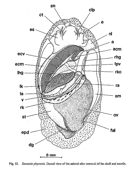
Pallial Complex: Pallial cavity half as deep as postcephalic visceral region, containing left ctenidium with osphradium, paired kidney openings, anus, and paired hypobranchial glands (Fig. 55).
Ctenidium bipectinate, crossing pallial cavity obliquely from posterior left to anterior right side. Efferent side connected to shell muscle by long efferent membrane (ecm); afferent side suspended from mantle skirt by short afferent membrane (acm).
Ctenidial lamellae dorsoventrally depressed. Ventral lamellae wider than dorsal lamellae. Right edge of ventral lamellae partly extended, visible in dorsal view (Fig. 55). Both lamellae provided with row of bursicles near efferent ctenidial axis. Lamellae supported by skeletal rods on efferent side. Afferent membran penetrated by transverse and anterior pallial veins (tpv, apv). Efferent axis of anterior free portion covered by osphradium.
Left hypobranchial gland (lhg) lying on mantle skirt in right portion of ctenidium, intersected dorsally by transverse pallial vessel. Right hypobranchial gland (rhg) attaching to right side of pallial section of rectum, greatly reduced into narrow strip.
Digestive System: Composition of oral, odontophoral, and radular muscles as those in Chlorostoma.
Postdorsal buccal tensor (pdt) and dorsal buccal tensors (dbt) present. Postmedian retractors (prs) and thin tensor muscle (trs) inserting on ventral side of radular sac. Jaws paired in oral tube. Anterior margin simple. Odontophoral cartilages consisting of longer anterior and shorter posterior pairs, united by ventral and oute approximator muscles.
Radular sac bifurcated at posterior end. Radular formula n-5-1-5-n. Tooth row symmetrical. Central tooth with short shaft, distinctly serrated cusp, and base forming broad and rounded lateral extensions. Inner four laterals with broad cusps with outer serration and enlarged bases with smooth outward extension. Outermost laterals similar to inner teeth in cusp morphology, but shaft straight without extension. Bases of all laterals overlapping. Protolateromarginal plates smooth, well-developed. Marginal teeth elongated with deep food groove below serrated cusps.
Sublingual pouch shallow, simple. Licker smooth. Radular diverticulum present. Salivary glands over buccal pouch, opening though longitudinal slits. Esophageal pouches (ep) enlarged on posterior side of buccal mass. Inner wall of mid-esophagus (me) papillate; dorsal and ventral folds twisted counterclockwise interiorly. Mid-esophagus constricted, followed by posterior esophagus (pe). Stomach (st) well-inflated, un-folded (Fig. 56 a); esophagus and ducts of digestive glands opening on right posterior side (Fig. 56 b); posterior apex giving rise to spiral gastric caecum (gc) with more than three volutions. Tooth of gastric shield on dorsal side of esophageal opening. Sorting area (sa) well-developed ventrally. Major and minor typhlosoles (mt, nt) continuing from posterior part of stomach into intestine. Intestine (i) bent four times to form two anterior loops on left side of mid-esophagus; rectum penetrating pericardium and ventricle; anus (a) opening beside right anterior rim of shell muscle.

Circulatory System: Pericardium extending along border of pallial cavity and visceral hump between paired kidneys (Fig. 55). Heart consisting of asymmetrically paired auricles and median ventricle. Right auricle (ra) extremely elongated along pericardial wall, connected to efferent pallial vessel through narrow part of anterior end. Outlet to ventricle opening from left anterior side. Left auricle (la) more developed than right one. Irregular margin of filtration chamber well-developed. Connection with ventricle arising from right side. Ventricle (v) greatly elongated throughout pericardium. Aorta bifurcating into anterior and posterior aortae without forming distinct bulbous aorta at their dividing point. Transverse pallial vessel (tpv) very clearly crossing left hypobranchial gland from right kidney to afferent ctenidial vessel.
Excretory System: Excretory system consisting of two kidneys (Fig. 55). Right kidney (rk) narrowly elongated along posterior margin of pericardium, extending ventrally to right anterior visceral region; lumen spacious; lamellate wall irregularly fenestrated. Right kidney opening (rko) projecting as short papilla. Left kidney (lk) lying within pallial cavity; lumen densely filled with long spicular papillae (Fig. 55). Left kidney opening simple short slit.
Reproductive System: Gonochoristic gonad lying on dorsal side of digestive glands (Fig. 55). At maturity, gonad also extending over stomach. In dissected specimens, gonoduct not distinct, but well developed gonad directly reaching anterior section of right kidney. Urogenital papilla not well differentiated in female.
Nervous System: Circumesophageal nerve ring hypoathroid. Cerebral commissure running over dorsal food channel. Labial ganglia and commissure absent. Visceral loop arising from right and left pleural ganglia. Statocysts on anterodorsal margin of pedal ganglia.
Broderipia iridescens (Broderip, 1834)
(Figs. 57 a-b, 58 a-f, 59 a-d)
Protoconch: Protoconch trochispiral, longitudinally elongate, well-inflated (Figs. 59 b-c). Surface of protoconch covered by characteristic reticulation, divided into four distinct zones in apical area. Lateral pouch of apical side with fine reticulate ridge sculpture.

External Anatomy: Soft parts completely limpet-form without spiral visceral hump. Mantle margin lacking specific sensory tentacles. Shell muscle (sm) horseshoe-shaped, not divided into bundles.
Head with snout (sn) and pair of papillate cephalic tentacles (ct), lacking cephalic lappets between ce
phalic tentacles. Outer lip of mouth thickened into oral disk, without microtentacles (Fig. 58 a). Surface of snout weakly papillate. Neck region with inhalant (left) and exhalant (right) neck lobes (nl), both slightly papillate with ciliary crown (Fig. 58 b). Eyes (e) open at tip of eyestalks.
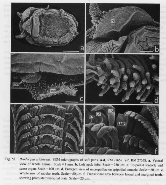
Foot sole marked by weak mid-ventral cleft. Epipodium in pallial groove provided with three pairs of epipodial tentacles (ept): two lateral, one posterior near midline (Fig. 58 a). Cephalic and epipodial tentacles bearing dense rows of ciliated microtentacles (Fig. 58 d). Base of each epipodial tentacle with large, knob-shaped epipodial sense organ (eso) (Fig. 58 c). Foot autotomy line and operculum absent.
Pallial Complex: Pallial cavity reaching one-half body length, containing left ctenidium with osphra-dium, paired kidney openings, and paired hypobranchial glands (Fig. 57 a).

Ctenidium bipectinate, dorsoventrally depressed. Efferent ctenidial membrane (ecm) barely evident along inner side of shell muscle. Afferent membrane reduced. Dorsal ctenidial lamellae narrower than ven
tral lamellae, so that extended part of ventral lamellae visible on right side. Both lamellae with bursicles on efferent side. Lamellae and efferent free axis supported by skeletal rods. Osphradium lying on efferent free axis.
Hypobranchial glands paired, not well-developed. Right gland (rhg) greatly reduced, situated on right side of rectum near anus. Left gland (lhg) larger than right, lying far removed from right gland on pallial roof, contacting left side of rectum and anterior margin of left kidney posteriorly.
Digestive System: Composition of oral, odontophoral, and radular muscles identical to that in Chloros-toma. Postdorsal buccal tensor and dorsal buccal tensors present. Postmedian retractors and tensor muscle inserting on ventral side of radular sac.
Jaws divided into paired plates, distinguished by dark color on oral tube, anterior margin smooth. Odontophoral cartilages consisting of elongated anterior and nodular small posterior pairs connected with ventral and outer approximators.
Radular sac with clearly bifurcated posterior end. Radular formula n-5-1-5-n (Figs. 58 e, f). Tooth row symmetrical. Central tooth with short, constricted shaft below well-developed cusp; base with triangular lateral projections. Lateral teeth with cusps similar to that of central, becoming prominent toward outermost laterals. Bases of inner four laterals overlapping with basal extensions; that of outermost teeth smooth. Protolateromarginal plates (LMP) broadly developed, originating from base of innermost marginals. Marginals elongated with distinct cusps.
Sublingual pouch lacking glandular outgrowth, very shallow. Licker small and smooth. Radular diver-ticulum present. Salivary glands (sg) without ducts, lying on dorsolateral sides of buccal cavity, partly over-lapping dorsal food channel of anterior esophagus (Fig. 57 b). Mid-esophagus (me) marked by enlargement of esophageal pouches, twisting counterclockwise, terminating in constriction. Posterior esophagus (pe) straight, extending posteriorly on floor of visceral mass.
Stomach (st) pyriform, lying on left posterior side of visceral mass (Fig. 57 b). Posterior esophagus and ducts of digestive glands opening on right posterior side, intestine on left anterior end. Posterior apex of stomach with non-spiral, Small gastric caecum. Interior of stomach clearly defined by sorting area, gastric shield, tooth of gastric shield, and intestinal groove between major and minor typhlosoles.
Intestine (i) coiling only twice, generating single anterior loop along right side of mid-esophagus. Rectum penetrating pericardium and ventricle. Anus (a) opening near anterior right end of shell muscle.
Circulatory System: Pericardium forming part of visceral hump, contacting posterior limit of pallial cavity behind left kidney (Fig. 57 a). Heart consisting of right and left auricles (ra, la) and single ventricle (v), all markedly transversely elongated.
Excretory System: Two kidneys facing each other on each side of rectum (Fig. 57 a). Right kidney enlarged, composed of two clearly separated sections. Posterior lobe (prk) hollow, with smooth interior surface; gonoduct opening posteriorly. Anterior lobe (ark) lamellate internally, opening ventrally. Anterior and posterior lobes connected to each other through narrow canal. Right renopericardial duct opening into peri-cardium beneath right auricle. Opening of right kidney slightly papillate. Left kidney (lk) composed of papillary sac. Left renopericardial duct opening in median part of anterior pericardial wall. Opening of left kidney slit-like.
Reproductive System: Gonad (g) spreading over posterior dorsal surface in both sexes. Gonoduct opening into central part of posterior lobe of right kidney (Fig. 57 a).
Nervous System: Circumesophageal nerve ring hypoathroid. Pedal and pleural ganglia markedly con-centrated. Cerebral commissure running over anterior part of dorsal food channel. Labial ganglia and commissure absent. Visceral loop arising from right and left pleural ganglia. Pedal cords scalariform. Statocysts attached to anterior sides of pedal ganglia.
Cantharidus sp. cf. callichroa (Philippi, 1850)
(Figs. 59 e-h)
Protoconch: Protoconch nearly discoidal, inflated, paucispiral. Coiling direction orthos trophic. Lateral pouch on apical side projecting to apertural side, with extension producing clear suture as deep cleft. Pouch on basal side projecting, marking distinct pouch-like region. Surface on apical side ornamented by irregularly scattered crystal deposits coalescing to form several spiral threads becoming gradually convergent adaperturally. Protoconch-teleoconch boundary marked by thickened axial lip.
Calliostoma sp.
(Figs. 60 a-d)
Protoconch: Protoconch longitudinally elongated, well-inflated. Adapertural part tightly coiled to orthostrophic direction with distinct suture line. Lateral pouch clearly visible on apical side. Protoconch forming distinct angle to teleoconch, exhibiting heterostrophic appearance. Ornament on protoconch represented by wholly regular hexagonal network composed of vertically aligned columnar prismatic crystals superim-posed vertically on smooth base. Boundary between protoconch and early teleoconch distinct, with thickened axial ridge.

Lirularia? minima (Golikov in Golikov and Scarlato, 1967)
(Figs. 60 e-h)
Protoconch: Protoconch initially planispiral, becoming slightly hyperstrophic in final stage, so that protoconch situated at quite different orientation against axis of orthostrophic, conispiral teleoconch. Surface covered by sharp net-like sculpture; some spiral threads visible in ventral view. Protoconch-teleoconch boundary marked by broad axial band.
Family Skeneidae Clark, 1851
Pondorbis japonicus Ando and Habe, 1980
(Figs. 61 a-d)
Protoconch: Protoconch longitudinally elongated, strongly distorted, heavily heterostrophic. Apex pointed ventrally; ventral side of protoconch well inflated. Sculpture almost homogeneously pitted or with polygonal network sculpture. Ridges partially emphasized. Protoconch-teleoconch boundary marked by axial ridge.
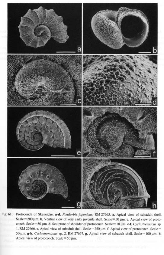
Cyclostremiscus sp. 1
(Figs. 61 e-f)
Protoconch: Protoconch almost planispiral, well-inflated. Lateral pouch not appearing differentiated with globular protoconch outline. Well-inflated abapertural part marking very clear sutural line as deeply cleft groove. Surface of median apical area mostly smooth, free from sculpture except for very minute vermicular deposit. Remaining area roughly sculptured by flat ridge with densely pitted depressions. Protoconch-teleoconch boundary marked by thick ribs that are completely continuous with spiral ribs of teleoconch.
Cyclostremiscus sp. 2
(Figs. 61 g-h)
Protoconch: Protoconch planispiral. Apical area almost flat. Lateral pouch on apical side small, not extended laterally. Suture of protoconch clear. Sculpture of protoconch continuous, consisting of widely radiating spiral ridges occurring only on apical side. Lateral pouch on apical side delimited by short spiral ridge
crossed by six or more fine oblique ribs. Apertural margin of protoconch very thick, inflected.
Family Lepetodrilidae McLean, 1988
Lepetodrilus nux (Okutani, Fujikura and Sasaki, 1993)
(Figs. 62,63 a-e, 64 a-d, 65 a-h)
External Anatomy: Soft parts limpet-shaped with apex projecting posteriorly. Mantle margin divided into two folds by deep periostracal groove (pg). Outer fold (of) thinner and smoother; inner fold (if) thicker, fringed by numerous microtentacles (Fig. 65 a).

Head with snout (sn), and pair of non-papillate cephalic tentacles (ct). Snout short; neck region prolonged forward. Outer lip of mouth smooth, without sensory projection, lacking oral lappets. Posterior bases of cephalic tentacles extended laterally; eyes not located in serial sections. Neck lobes and cephalic lappets absent. Penis in male not confirmed (material examined all female).
Anterior margin of ventral foot sole demarcated by groove marking opening of anterior pedal gland. Non-papillate epipodial tentacles represented by long folds with three pairs of protrusions on sides of foot. Tuberculate epipodial sense organs at pointed tip of epipodial tentacles. Operculum absent.
Paired shell muscles (sm) horseshoe-shaped, posteriorly connected to filamentous muscles below visceral hump, not divided into bundles or penetrated by blood vessels (Fig. 62).
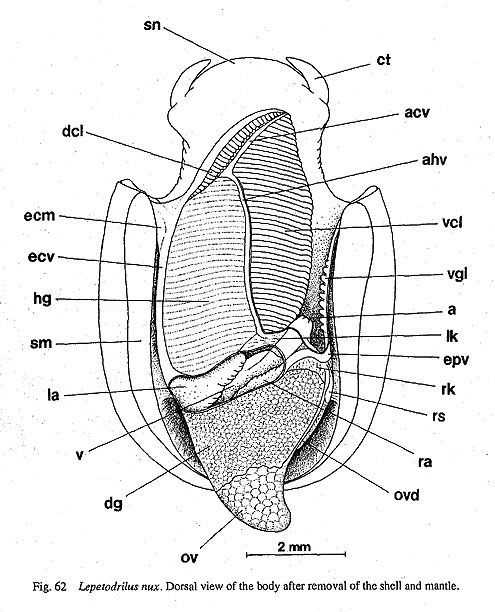
Pallial Complex: Pallial cavity attaining two-thirds of body length, containing left ctenidium, left osphradium, gill leaflets on right side, paired kidney openings, anus, and left hypobranchial gland.
Ctenidium greatly modified from typical bipectinate ctenidium. Lamellae bipectinate in anterior third but monopectinate in remaining posterior area. Ctenidial axes not supported by membrane, directly attached to mantle skirt in monopectinate area. In bipectinate part, dorsal lamellae (dcl) very short, restricted to narrow outer crescent-shaped zone of bipectinate area (Figs. 62, 65 b). Ventral lamellae (vcl) dorsoventrally depressed, fused with base of hypobranchial gland (Fig. 62). Respective ventral lamellae elongated feather-shaped. Each ctenidial axis containing paired muscles and median nerve. Each leaflet with frontal, abfrontal, lateral, and terminal cilia; efferent side internally supported by paired skeletal rods; leaflets with series of
shallow grooves near apex in ventral view (Fig. 65 c). [These cannot be identified as bursicles, because of absence of interiorly ciliated lumen.]
Right side of pallial cavity lacking true ctenidium. Nine to twelve gill leaflets projecting from efferent pallial vessel (epv) which runs along interior shell muscle (Fig. 62). Surface entirely covered with cilia that appear weakly organized into zones (Fig. 65 d).
Osphradium not distinguishable in superficial observation, but histological thin sections revealed darkstained sensory epithelium covering free portion of efferent ctenidial axis, extending along anterior end of left shell muscle. Osphradial epithelium including tall cells with elongated nuclei. Osphradium not located on right side.
Well-developed hypobranchial gland (hg) widely attached to left half of mantle skirt, partitioned into narrow transverse sections by vessels from afferent to efferent hypobranchial vessels (Fig. 62). Venous blood transported from transverse pallial vessel to afferent vessels of ctenidial lamellae through hypobranchial glands. Therefore, intervals of sectors of hypobranchial glands completely corresponding to those of vessel of ctenidial lamellae.
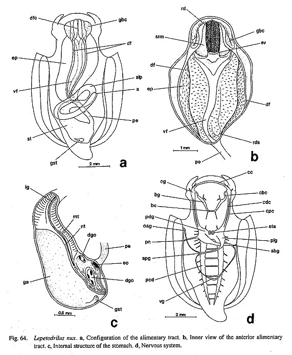
Digestive System: Oral tube surrounded by circular buccal sphincter and constrictor (bs, bct). Buccal tensors (bt) extending from ventral side of sublingual pouch to wall of snout between inner ventral protractors and lateral protractors. Mandible protractors (mp) arising from surface of buccal constrictor, and mandibular retractors (mr) appearing from inside of buccal sphincter. Retractors of esophageal valve (rov) inserted on dorsal side of radular sac, covered by postdorsal buccal tensor (pdt).
Odontophores fixed by lateral protractor, posterior depressor, anterior levator, ventral protractor muscles (Figs. 63 a, b). (1) Lateral protractors (lp) arising from posterior cartilages, running along sides of odontophore, attaching dorsolaterally to buccal constrictor. (2) Posterior depressors (pd) inserted near outer lateral comers of posterior cartilages, crossing over postdorsal buccal tensor (pdt), directed downward, united with floor of body cavity. (3) Anterior levators (al) occurring in anterolateral extensions of anterior cartilages, ascending along cartilages on inner sides of lateral protractors and dorsal buccal tensors, emerging dorsally just beside buccal cavity. (4) Inner and outer ventral protractors (ovp, ivp) inserted on ventral surface of posterior cartilages, extending to sublingual pouch.

Left and right halves of odontophore united by two kinds of tensors. (1) Postdorsal buccal tensor (pdt) originating from sides of posterior cartilages under insertions of posterior depressors. (2) Dorsal buccal tensors (dbt) consisting of two parts: dorsal part extending over lateral protractor; outer ventral part running downward, fusing with lateral margin of subradular membrane.
A pair of dark brownish jaws (j) attached to dorsal walls of oral tube at posterior two-third. Finely split, rod-bearing anterior edge exposed to opening of mouth (Figs. 63 b, e).
Radutar sac short, straight; posterior end deeply bifurcated (Fig. 64 b). Radular formula 25+-5-1-5-25 +(Fig. 65 e). Tooth row asymmetrical. Central tooth with bilateral basal extensions overlaid on bases of innermost lateral teeth; cusp longitudinally rectangular with very minute serration of denticles. Five pairs of lateral teeth of almost even width; cusps fringed by fine serrated denticulation; shafts almost straight. Base of outer teeth interlocked by inner teeth. Cusps of innermost lateral teeth longer than those of second to fifth teeth, showing steep V-shaped arrangement. Lateromarginal plates absent. Marginal teeth with broadly elongated shafts.
Subradular membrane attached by paired thin median protractors (mpr) and thick lateral protractors (lpr) (Fig. 63 b). Lateral protractors terminating in origin on posterior cartilages. Median protractors elongated along radular sac, anchored on pedal musculature below mid-esophagus.
Retractors of radular sac extending from origin on inner sides of posterior cartilages to insertion on ventral mid-part of radula between anterior cartilages. Tensor of radular sac (trs) as thick as median protractor muscles (Fig. 63 b), arising from underside of radular sac just behind posterior end of buccal mass, running straight forward along mid-line, bifurcated anteriorly into symmetrical strands, fixed to floor of sublingual pouch, crossing over median and lateral protractors of subradular membrane. Postmedian retractors of radular sac absent.
Buccal mass containing single pair of odontophoral cartilages (ca), connected by ventral and outer approximator muscles (vap, oap) (Figs. 63 c, d).
Sublingual pouch (slp) shallow, lacking glandular outgrowth (Fig. 63 a). Licker (lic) on anterior tip of subradular membrane elongated, smooth (Fig. 63 b). Salivary glands totally absent around buccal cavity, but sides of buccal cavity lined by tall glandular cells.
Beginning of esophagus marked by esophageal valve (ev), separated from deep radular diverticulum. Anterior esophagus extended laterally to form large esophageal pouches of mid-esophagus, remarkably dilated laterally, also extended ventrally to completely enclose posterior part of radular sac (Fig. 64 b). Interior of esophagus provided with paired longitudinal folds (Figs. 64 b, 65 f). Dorsal folds (df) paired throughout anterior to mid-esophagus. Ventral folds (vf) beginning from sides of esophageal valve, converging to fuse into thick single fold, extending almost straight back, only posterior part twisted counterclockwise, Inner surface of mid-esophagus (me) wholly covered with papillate projections of esophageal gland, except in ciliated dorsal food channel (dfc) (Fig. 65 f). Posterior esophagus (pe) greatly narrowed, obliquely postured on right side.
Stomach (st) pyriform, on bottom of visceral mass (Fig. 64 a). Paired ducts from digestive glands entering near opening of esophagus (eo). Posterior apex of stomach weakly projecting as small gastric caecum. Internally, most area of left side occupied by smooth cuticularized gastric shield (gs) (Fig. 64 c). Tooth of gastric shield (gst) projecting on posterior wall of stomach. Right side narrowly sculptured by folds; sorting area not well-developed. In posterior half, intestinal groove (ig) deeply formed between major and minor typhlosoles.
Intestine folded twice to form single anterior loop (alp) on posterior right side of mid-esophagus (Fig. 64 d). Rectum penetrating pericardium and ventricle (v). Anus (a) opening into posterior right corner of pallial cavity.
Circulatory System: Heart consisting of right and left auricles and single ventricle (Fig. 62). Right auricle (ra) smaller than left (la). Initial part of aorta not enlarged into bulbous aorta.
Efferent pallial vessel (epv) on right side draining into pallial cavity along shell muscle, running over right kidney, finally entering right auricle. Circulation in left pallial cavity beginning from transverse pallial vein arising from anterior margin of left kidney, soon connecting to afferent hypobranchial vein (ahv). Branches from the vessel supplying hypobranchial gland, entering afferent side of ctenidium, finally joining efferent ctenidial vessel (ecv). Therefore, blood necessarily entering ctenidium via hypobranchial gland. Afferent hypobranchial vessel extending anteriorly along right margin of hypobranchial gland, becoming afferent ctenidial vessel (acv) at termination of hypobranchial gland. Efferent ctenidial vessel extending from free tip to entire length of ctenidium. At anterior base of efferent ctenidial membrane, left efferent pallial vessel joining efferent ctenidial vessel, entering left auricle.
Excretory System: Excretory system consisting of right and left kidneys (Fig. 62). Right kidney (rk) extending to dorsal surface of visceral mass, also continuing ventrally. Oviduct in female discharged within dorsal lobe. Opening of right kidney (rko) slit-like.
Left kidney (lk) overhanging in pallial cavity, receiving blood from blood space below kidney. Left kidney opening into pallial cavity ventrally through minute slit-like opening. Renopericardial duct present on both right and left sides.
Reproductive System: Specimens studied all females. Ovary (ov) lying over digestive glands in pointed visceral hump under shell apex (Fig. 62). Thin oviduct extending from right side of ovary, passing over exterior surface of visceral mass. Oviduct connected to kidney though short pathway of renopericardial canal.
Ova discharged into pallial cavity through right kidney opening. Floor of pallial cavity on right side lined with tall ciliated cells, perhaps serving for transportation of gametes.
Right pallial wall deeply excavated outside of right kidney into deeply elongated sac that acts as receptaculum seminis (rs) (confirmed by optical and scanning electron microscopy of serial sections that this structure fills with spermatozoa) (Figs. 65 g, h).
Nervous System: Circumesophageal nerve ring hypoathroid (Fig. 64 d). Cerebral ganglia at bases of cephalic tentacles; pleural and pedal ganglia (plg, pdg) tightly fused behind buccal mass. Cerebral commissure (cc) running in front of jaw plates. Cerebropleural and cerebropedal connectives (cdc, cpc) very long, correlated with elongation of neck region.
Several pairs of labial nerves extending from ventral part of cerebral ganglia but not fusing to form labial commissure. Labial ganglia absent. Cerebrobuccal connectives (cbc) arising from inner ventral sides of cerebral ganglia.
Visceral loop originating from both right and left pleural ganglia. In spite of great depth of pallial cavity, visceral loop not very long because of posterior location of pleuro-pedal ganglia. Connectives from right and left pleurals to subesophageal and supraesophageal ganglia (sbg, spg) rather thick; posterior connectives becoming extremely thin toward visceral ganglion beneath left kidney.
Left pleural ganglia laterally providing nerves to pallial, parietal, and pedal regions. Ctenidia innervated by nerve from supraesophageal ganglion. Corresponding nerve on right side extending toward shell muscle. Osphradial ganglion (osg) only on left side. Right side of equivalent nerve and ganglion reduced. Zeugoneury not confirmed.
Pedal ganglia extending backward to thick pedal nerve cords. Pedal commissure well-developed, forming scalariform pedal nerve system. Statocysts (sta) attached to inner anterodorsal sides of pedal ganglia.
|