Family Phenacolepadidae Pilsbry, 1895
Cinnalepeta pulchella (Lischke, 1871)
(Figs. 81 a-b, 82 a-b, 83 a-c, 84 a-b, 85 a-f)
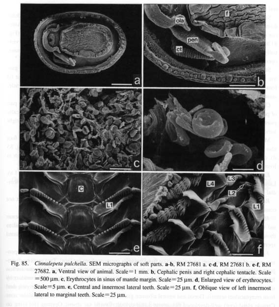
External Anatomy: Animal limpet-shaped. Mantle margin clearly divided into inner and outer folds by deep periostracal groove. Inner fold of mantle margin with mamillate microtentacles (Figs. 85 a, b). Head with short snout (sn), pair of long non-papillate cephalic tentacles (ct), and short eyestalks (es) at outer bases (Fig. 81 a). Outer lip of mouth smooth, extended laterally with oral lappets. Eyes (e) closed on eye-stalks. In males, long tentacular penis (pen) arising from ventral side of base of right cephalic tentacle (Fig. 85 b). Epipodium lacking epipodial tentacles and sense organs. No trace of internal operculum located in pedal musculature in serial sections. Shell muscle horseshoe-shaped without division or penetration by blood vessels.
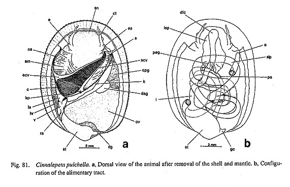
Pallial Complex: Pallial cavity deepened in anterior two-thirds of body, containing left ctenidium, left kidney opening, anus, and genital opening(s) (Fig. 81 a). Hypobranchial gland and "vestigial gill" absent on right side.
Ctenidium (c) bipectinate. Greater portion of ctenidial axes lacking ctenidial membranes. Internal struc-ture of axes and surface cilia identical to those in Nerita. Ctenidial lamellae depressed, with centrally pro-jecting terminal ridges. Efferent side lacking bursicles and skeletal rods. Osphradium (os) attached to inner margin of shell muscle, provided with lateral ciliated zones.
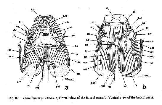
Digestive System: Odontophore connected to body wall by median and posterior levators (ml, dl), poste-rior depressors (pd), anterior levators (al), lateral protractors (lp), ventral protractors (ovp, ivp), and dorsal
buccal Censors (dbt), (Figs. 82 a, b). Postdorsal buccal tensor (pdt) connecting right and left sides of poste-rior cartilages.
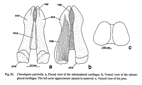
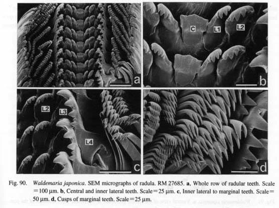
Jaws consisting of paired thin plates attached to dorsal side of oral tube (Fig. 83 c). Radular sac almost straight, not coiled. Posterior end simple. Radular formula n-(1+3)-1-(3+1)-n (Figs. 85 e, f). Tooth row symmetrical. Central tooth nearly square in frontal view, with slightly extended basal margins, without dis-tinct cusp. First inner lateral teeth greatly obliquely elongated; outer comer situated more anteriorly than inner corner; cusp saw-like, with fifteen sharp denticles; base with reflected flaps projecting as sharp median denticle. Second inner laterals small, almost fused with first teeth. Third laterals enrolled inside, with poste-riorly elongated base. Outer (fourth) laterals longitudinally straight; cusp angulate with three weak denticles. Lateromarginal plates absent. Marginal teeth thin, long, with sharply denticulate cusps.
Subradular membrane controlled anteriorly by median and lateral protractors (mpr, Ipr), and posteriorly by retractors (rsr). Radular sac controlled by retractor muscles (rrs) between pairs of cartilages. Tensor and other retractors absent in radular sac.
Odontophore cartilages consisting of longer anterior and smaller posterior pairs of cartilages (aca, pca) and unpaired median cartilage (mca) (Figs. 83 a-b). Paired anterior cartilages united by ventral approximator (vap); anterior and posterior cartilages connected by outer approximators (oap). Tensor muscles of anterior cartilage (tac) originating from body wall, inserting on sides of anterior cartilages, passing between lat
eral protractors (lp) (Fig. 82 b). Sublingual cartilage absent.
Sublingual pouch (slp) deep, with outgrowth of sublingual gland (slg) on each side (Fig. 82 b). Radular diverticulum shallow. Licker absent at anterior part of subradular membrane. Salivary glands absent. Anterior esophagus accompanied by long lateral pouches (lep) in septate posterior esophageal glands (peg) (Fig. 81 b). Terminal section of glands posteriorly separated from main channel of esophagus, extending ven-trally along posterior slope of buccal mass. Posterior esophagus (pe) running along base of visceral mass, opening into stomach on right side.
Stomach (st) provided with pair of openings to digestive glands, small gastric caecum (gc), toothed gastric shield, narrow sorting area, and intestinal groove between major and minor typhlosoles. Intestine (i) forming complicated course of about five loops (Fig. 81 b); rectum penetrating pericardium and ventricle, anus (a) opening beside right end of shell muscle.
Circulatory System: Pericardium compressed along inner wall of shell muscle on left side. Posterior part of pericardium extending into ventral part of stomach as narrow tube. Heart consisting of asymmetrically paired auricles and single ventricle. Left auricle (la) connected to efferent ctenidial vessel; reduced right auricle (ra) apparently lacking circulatory function.
Mantle margin thickened to develop into spongy space as respiratory surface (Fig. 85 c). Side of foot also with particular blood space. Discoidal erythrocytes, each 8 µm in diameter and 1.6 µm thick (Fig. 85 d), densely distributed throughout vascular system. Because of their presence, animal entirely reddish in living condition, immediately turning pale after fixation.
Excretory System: Single left kidney (k) in elongated area of pallial cavity wall along rectum (Fig. 81 a). Lumen divided into glandular and non-glandular areas. Kidney opening (ko) below origin of afferent ctenidial vessel as non-papillate, simple, longitudinal slit.
Reproductive System: Gonochoristic gonad (g) lying over digestive glands and stomach. Gonoduct not connected to kidney, exhibiting sexual dimorphism.
Male Gonoduct (Fig. 84 a): Vas deferens (vd) from ventral side of testis coiling complexly to form seminal vesicle (sv), leading anteriorly to prostate (pr) covered by another glands histologically identical to those of annex gland in Nerita. Basal gland absent. Opening (mo) lying at ventral tip of gonoduct. Ciliated sperm groove between gonopore and cephalic penis absent.
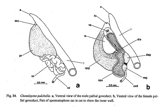
Female Gonoduct (Fig. 84 b): Female gonoduct with two genital openings (diaulic). Vagina (va) opening on top of anterior section; posterior section functioning as spermatophore sac (sps). Two sections distin-guished externally by constrictions and internally by corrugated or papillate sculpture. Anterior section leading through narrow slit into neighboring receptaculum seminis (rs), which extends to pallial oviduct by short duet. Oviduct connected to ventral albumen gland (vag); dorsal albumen gland (dag) receiving duct from receptaculum seminis. Dorsal and ventral albumen glands separated by distinct median glandular zone. Capsule gland (cpg) well-developed throughout pallial oviduct. Crystal sac absent. Opening (fo) lying on an terodorsal side of vagina.
Nervous System: Circumesophageal nerve ring hypoathroid. Pedal and pleural ganglia concentrated. Right and left pleural ganglia connected by pleural commissures. Cerebral ganglia (cg) ventrally united by labial commissure (lc) without labial ganglia (Fig. 81 b). Connectives to buccal ganglia originating in labial commissure, not in cerebral ganglia. Supra- and subesophageal portions of visceral loop both arising from pleural ganglia. Viscero-supraesophageal connective not clearly traceable. Pedal cords apparently lacking distinct commissures. Statocysts on dorsal sides of pedal ganglia below pleural commissure.
Phenacolepas sp.
(Figs.86 a-d)
Protoconch: Protoconch globular, multispiral. Apex projecting to right due to orthostrophic growth. Inner side of protoconch completely absorbed (Fig. 86 d); exterior part of protoconch including coiled suture retained in shell apex.

Family Hydrocenidae Troschel, 1856
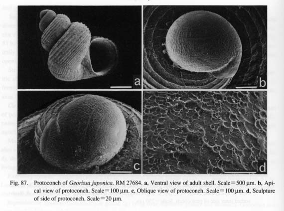
Georissa japonica Pilsbry, 1900
(Figs. 87 a-d)
Protoconch: Protoconch completely globular. Narrow aperture formed anteroventrally, generating short deep suture. Surface covered with irregular depressions in addition to circular lines. Protoconch-teleoconch boundary expressed as smoothly thickened lip.
Family Helicinidae Féussac, 1882
Waldemaria japonica (A. Adams, 1861)
(Figs. 88 a-b, 89 a-e, 90 a-d, 91 a-b)
External Anatomy: Animal discoidal, lacking spiral visceral hump, although shell spirally coiled. Axial cleft formed for columellar septum (Figs. 88 a, b).
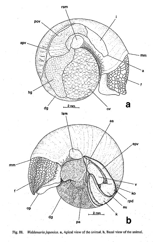
Mantle margin lacking circumpallial tentacles. Head with short snout, non-papillate cephalic tentacles, eyestalks. Outer lip of mouth simple, without oral lappets or microtentacles. Eyes lying on eyestalks outside bases of cephalic tentacles, closed by cornea. Male lacking cephalic penis on right side.
Foot rather clearly divided into lateral and ventral portions in fixed specimens. Ventral part smooth, with opercular attachment (op). Surface of dorsal part with rough appearance. Surface of epipodium tuberculate, lacking sensory projections. Calcareous operculum present.
Shell muscles paired, completely divided into dorsal (right) and ventral (left) portions (rcm, lcm). Mus
cles not divided or penetrated by blood vessels. Head retractor muscle indistinct.
Pallial Complex: Pallial cavity deep, attaining more than half of body whorl, containing kidney opening, pallial gonoduct, anus, and right hypobranchial gland (Figs. 88 a, b). Ctenidium absent, replaced by vascu-larized mantle skirt. Blood vessels densely developed, especially near efferent and afferent pallial vessels. Osphradium absent. Hypobranchial gland (hg) well-developed in deepest region of pallial cavity roof, dis-charging into pallial cavity via short hypobranchial duct on posterior side of vaginal opening.
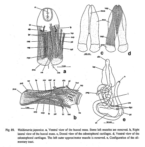
Digestive System: Oral tube surrounded by buccal sphincter and constrictor (bs, bct) with mandibular protractors and retractors (mp, mr). From ventrolateral sides near outer margin of jaws, thin buccal tensors (bt) extending ventrally to ventral body wall.
Muscle controlling odontophore including posterior depressors (pd), anterior levators (al), outer posterior levators (opl), lateral protractors (lp), and outer and inner ventral protractors (ovp, ivp) (Figs. 89 a, b). Median and dorsal levators and dorsal buccal tensor absent. Posterior part of cartilages united by postdorsal buccal tensor (pdt).
Jaws composed of simple translucent plates entirely adhering to dorsal side of oral tube. Inner surface and anterior margin not sculptured.
Radular sac almost straight, terminating near opening of esophagus to stomach. Posterior end simple without bifurcation. Radular formula n-(1+3)-1-(3+1)-n (Figs. 90 a-d). Radular row symmetrical. Central tooth longitudinally rectangular, with weak lateral extensions and simple plate-like cusp. First and second inner lateral teeth similar in size to central tooth; cusps serrated with four to six denticles. Third lateral teeth reduced, plate-like, situated at base of outer lateral teeth. Outer (fourth) lateral teeth greatly enlarged, exhibiting very characteristic complex form; cusp prolonged on inner anterior sides, where sharply serrated with five denticles; base strongly projecting backward in median part, also extending toward outside.
Lateromarginal plates absent. Marginal teeth with two to six acute denticles from inner to outer sides.
Muscles inserting on radular sac and subradular membrane consisting of median protractors (mpr), lat-eral protractors (lpr), retractors of subradular membrane (rsr), and retractors of radular sac (rrs) (Figs. 89 a, b). Postmedian retractors and tensor of radular sac absent.
Odontophore containing anterior, posterior, and median cartilages (Figs. 89 c, d). Anterior cartilages (aca) extremely elongated; posterior cartilages (pca) small. Pair of anterior cartilages connected by ventral approximator (vap). Anterior and posterior cartilages connected by outer approximator muscles (oap). Single rod-like median cartilage (mca) present between anterior cartilages above ventral approximator. Sublin-gual cartilage (as in Nerita) absent. Lateral extensions of anterior cartilages inserted by tensor muscles (tac) (Fig. 89 b). Anterior tensors ofodontophore (at) arising from more anterior part than it.
Sublingual pouch (slp) lacking particular outgrowths of sublingual gland (Fig. 89 a). Licker absent. Radular diverticulum definitely present. Salivary glands not developed as distinct glands outside buccal cavity. Anterior esophagus dorsoventrally depressed, accompanied by lateral esophageal pouches (lep) over buccal cavity region. Esophagus curving toward left; posterior section of esophageal pouches arising from it, forming extremely elongated posterior esophageal glands (peg) over intestinal loops (Fig. 89 e). Mid-esophagus not differentiated. Paired ducts from digestive glands connecting with stomach on posterior side. Stomach provided externally with short crescent-shaped gastric caecum (gc) and internally with gastric shield, tooth of gastric shield, sorting area, major and minor typhlosoles, and intestinal groove. Intestine (i) tightly coiling, forming several loops. Rectum passing kidney ventrally, but not passing through pericar-dium or ventricle. Anus (a) opening on anterior right side of pallial cavity together with genital opening.
Circulatory System: Pericardium situated on left half of posterior end of pallial cavity. Heart consisting of single auricle and single ventricle (Fig. 88 b). Auricle (au) triangular, attached to efferent pallial vessels widely. Ventricle (v) almost free except for narrow vascular connection. Within pericardium, aorta dividing into anterior and posterior aortae (aa, pa). Bulbous aorta not formed. Afferent pallial vein (apv) from kidney running along gonoducts. Efferent pallial vein running along pericardium, directly connecting with ventricle.
Excretory System: Single left kidney (k) lying on posterior side of pericardium (Fig. 88 b), internally divided into glandular part on anterior side and non-glandular part on posterior side. Distal part of kidney ex-tending to right of pericardium, opening into pallial cavity though small slit-like pore (ko). Renopericardial duct (rpd) extending left from distal region to pericardium.
Reproductive System: Gonochoristic gonad lying on dorsal surface of uncoiled visceral hump (Fig. 88 a). In both sexes, tissues of gonads aggregating in to cylindrical form, appearing as rounded patterns on dorsal surface. Therefore, sexes not distinguishable according to granulation of gonadal tissue. Gonoduct not con-nected with kidney, exhibiting sexual dimorphism.
Male Gonoduct: Male gonoduct developing into prostate within pallial cavity. Vas deferens entering
prostate from posteroventral end of pallial gonoduct. Seminal vesicle not developed in any section of vas deferens. Prostate divided into anterior and posterior sections by constriction. Anterior portion of prostate with long prostate pouch on dorsal surface. Annex gland and basal gland absent on pallial gonoduct. Gonopore opening in association with anus. External copulatory organ absent. Production of spermato-phores not found.
Female Gonoduct (Figs. 91 a, b): Female gonoduct with two genital openings (diaulic). Vaginal opening (va) lying in posterior end of pallial cavity, followed posteriorly by bursa copulatrix (bcp) which ap-proaches dorsal surface over pallial oviduct (pov) and is visible through mantle. Between vagina and bursa, "provaginal sac" (pvs) (Thompson, 1980) connected by way of narrow ducts; function not detected. Bursa copulatrix entering pallial oviduct from right side.
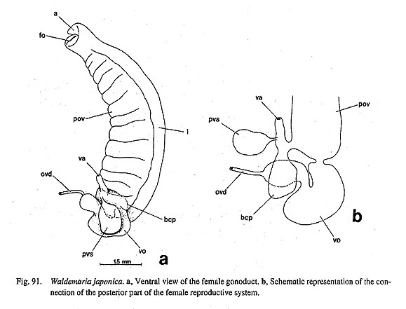
Oviducts from ovary expanding and twisting to form "V-organ" (vo) (Thompson, 1980). V-organ lying between dorsally sifted bursa copulatrix and ventrally positioned provaginal sac (Fig. 91 a). Distal part of V-organ communicating with pallial gonoducts on left side. From there pallial gonoducts running along rectum, terminating in anterior pallial cavity. Opening of pallial gonoducts incorporated with anus into common chamber.
Nervous System: Circumesophageal nerve ring hypoathroid. Cerebral ganglia at bases of cephalic tenta-cles. Cerebral commissure running in front of buccal cavity over oral tube. Pleural and pedal ganglia com-pletely fused behind buccal mass with long cerebropedal and cerebropleural connectives. Pleural ganglia connected by pleural commissure.
Cerebral ganglia united ventrally by labial commissure, and dorsally by cerebral commissure. Labial ganglia absent Buccal ganglia connected to labial commissure through labiobuccal connectives.
Visceral nerve loop arising only from right pleural ganglion. Thinner supraesophageal part of visceral loop originating more anterior than that of thicker subesophageal part. Supraesophageal and subesophageal ganglia weakly developed. Statocysts on dorsal sides of pedal ganglia, connected by short nerve.
|