Family Fissurellidae Fleming, 1822
Scutus (Aviscutum) sinensis (Blainville, 1825)
(Figs. 34,35 a-c, 36 a-f, 37 a-f)
External Anatomy: Mantle clearly divided into three folds, with outer fold always extending dorsally over shell margin. Anterior part of mantle bearing longitudinal mantle slit (Fig. 36 d), although its presence
hardly reflected in shell morphology. Mantle margin without circumpallial tentacles and long pallial tentacles.

Head with truncated snout, thick non-papillate cephalic tentacles (ct), and small eyestalks (es) (Fig. 34). Circumoral area thickened by smooth outer lip. Eyes on eyestalks closed by epithelium, with vitreous body inside (Fig. 37 a). Retina composed of two layers of tall cells (Fig. 37 b); inner cell layer darkly pigmented in black; outer layer unpigmented in histological sections. Oral and cephalic lappets and neck lobes not developed.

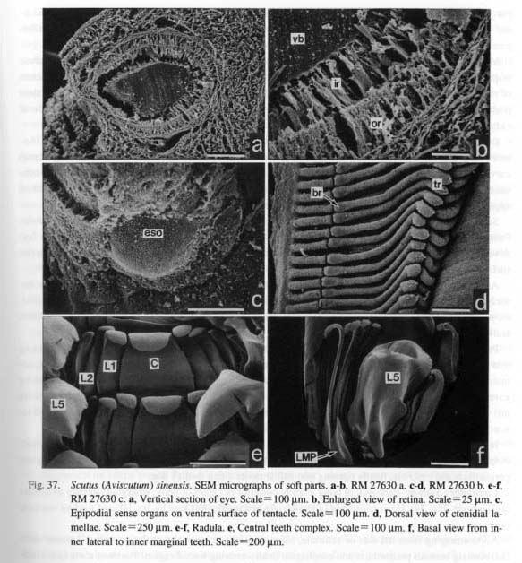
Epipodium with circle of triangular non-papillate epipodial tentacles. Epipodial sense organs (eso) lying on ventral surface of tentacles, not at base (Fig. 37 c). Operculum absent.
Shell muscle (sm) elongated horseshoe-shaped, not divided into bundles. Form of shell muscle asymmetrical by weak inward projection on left side of pericardium (Fig. 34). Pallial retractor muscle forming distinct attachment to shell. Head retractor muscle without independent attachment area.
Pallial Complex: Depth of pallial cavity reaching half length of area surrounded by shell muscle, including paired ctenidia with osphradia, anus, and kidney openings (Fig. 34). Hypobranchial glands absent.
Ctenidia (c) bipectinate, truly symmetrical in form and position. Ctenidial axes suspended on lateral pallial wall by efferent ctenidial membrane (ecm) and on roof of pallial cavity by afferent ctenidial membrane (acm). Each axis containing typical ctenidial structures of vessel, nerve, and retractor muscles. Lamellae triangular in outline, forming prominent terminal ridges (tr) projecting inside (Fig. 37 d). Efferent sides of lamellae with longitudinal array of bursicles (br) (Fig. 37 d). Surface of lamellae covered by three zones of ciliation. Lamellae supported by skeletal rods on efferent sides. Osphradia covering free portions of efferent axes.
Digestive System: Oral tube surrounded by sphincter and constrictor with mandibular protractors (mp)
and retractors (mr). From sides of oral tube, two distinct tensor muscles arising below layers of constrictors.
Muscles controlling orientation of odontophore comprising lateral protractors (lp), outer and inner ventral protractors (ovp, ivp), anterior levators (al), and posterior depressors (pd) (Figs, 35 a-c). Posterior levators absent. Posterior part of odontophore fixed by two kinds of tensor muscles. Dorsal buccal tensors (dbt) consisting of two distinct strands that fuse posteriorly. Inner strand originating from body wall, passing between lateral protractor and cerebral ganglion. Outer strand extending over inner strand, attaching to margin of subradular membrane. Postdorsal buccal tensor (pdt) inserting on both sides of posterior cartilages.

Jaws (j) wholly attached to inner wall of oral tube except fimbriate anterior tip (Fig. 35 b), divided into independent pair, but median zone connected by cuticularized membrane (Fig. 36 c).
Radular sac forming single loop to ventral side. Posterior end deeply bifurcated, connected to postdorsal buccal tensor (pdt) by thin muscle string (Fig. 35 a). Radular formula (n+1)-(1+4)-1-(4+1)-(1+n) (Figs. 37 e, f). Each teeth row almost symmetrical in central field, but outer lateral teeth asymmetrically interdigitated when radula closed by retraction. Central tooth trapezoidal, slightly asymmetrical, skewed to right side; cusp semicircular, lacking denticles; base also simple without lateral extension. Inner three pairs of inner laterals equal in length, but becoming more slender outside; cusps indistinct. Fourth teeth most slender among four paired inner laterals, concealed under inner margin of enlarged outer laterals. Outer (fifth) laterals sharply projecting with acutely pointed tip and subsidiary denticle at outer side of cusp; basal
part of shaft with thick ridge and lobate extension on each side. Lateromarginal plates strongly keeled; inner margin with close interaction with basal extension of outer laterals (Fig. 37 f). Marginals greatly elongated into filamentous form with finely serrated cusps.
Muscles controlling radular action consisting of median and lateral protractors of subradular membrane (mpr, lpr), retractors of subradular membrane (rsr), retractors of radular sac (rrs), and postmedian retractors of radular sac (prs) (Fig. 35 b). Tensor muscle of radular sac (trs) divided into right and left strands, their posterior ends attached to inner sides of postmedian retractors of radular sac (prs), not to ventral side of radular sac.
Odontophore containing two pairs of cartilages showing somewhat specialized form (Figs. 36 a, b). Anterior cartilages (aca) longitudinally elongated with weak anterolateral extensions. Posterior cartilages (pea) curving inside, attaching to posterior ends of anterior cartilages. Boundary between two cartilages connected by thin muscles dorsally, partially fused ventrally. Anterior cartilages ventrally inserted by ventral approximator (vap). Anterior and posterior pairs united by outer approximator muscles (oap).
Sublingual pouch shallow. Licker simple. Radular diverticulum present. No caecum (as in Diodora; Fretter and Graham, 1962: fig. 96, pdm) found between paired odontophoral cartilages. Salivary glands (sg) developed over buccal cavity (Fig. 36 d); opening not through ducts, but through longitudinal slits; dorsal urface ramified into vermiform outgrowths.
Anterior esophagus dilated to form esophageal pouches (ep), covering posterior end of buccal mass entirely. Dorsal folds paired along length of mid-esophagus; ventral folds fused in middle part. Wall of mid-esophagus extensively papillate. End of mid-esophagus marked by completion of twisting of dorsal folds and by disappearance of glandular papillae. Posterior esophagus (pe) very short.
Proximal part of stomach (st) well-inflated; outline pyriform (Figs. 36 d, e). Digestive glands opening into stomach through three large pores (dgo); two lying near opening of esophagus on posterior side, remaining one on right side. Gastric caecum (gc) small, crescent-shaped. Dorsal inner surface covered by gastric shield. Tooth of gastric shield (gst) projecting on posterior side. Sorting area (sa) and typhlosoles (nt, mt) well developed on ventral inner surface. Distal part of stomach and initial part of intestine elongated toward anterior left side, projecting over mid-esophagus.
Intestine (i) running along stomach posteriorly, forming anterior loop (alp) on right side of midesophagus (Fig. 36 d). Posterior part of intestine making another smaller loop above stomach, penetrating pericardium and ventricle, finally opening into pallial cavity along median line.
Circulatory System: Pericardium lying slightly posterior of center of body, enlarging laterally to contact shell muscle (Fig. 34). Heart consisting of larger right and smaller left auricles (ra, la), and median ventricle (v).
Aorta emerging from left side of ventricle, bifurcating into anterior and posterior aortae. Anterior aorta (aa) running beneath pericardium and esophagus, finally entering buccal region. Posterior aorta (pa) dividing into three main vessels. Right branch extending along posterior margin of right kidney, reaching gonad. Middle branch extending posteriorly, supplying blood mainly to stomach. Left branch penetrating into digestive glands.
Venous system around digestive organs well-developed, connected to kidneys. In front of kidneys, efferent renal vessels expanding to form small basibranchial sinus. Blood then flowing from afferent to efferent ctenidial vessels (acv, ecv), returning to right and left auricles.
Excretory System: Two extremely unequal kidneys (Fig. 34). Enlarged right kidney (rk) lying under pericardium, extending to dorsal surface on right posterior side of pericardium, also ventrally spreading over mid-esophagus; inner wall comprising of folded lamellae with irregular perforation. Renopericardial canal arising from right side, opening into pericardium near excretory pore of right kidney. Right kidney opening into pallial cavity via large papilla with longitudinal slit.
Left kidney (lk) greatly reduced, restricted to narrow space between posterior wall of pallial cavity and left auricle. Left kidney opening simple small pore without papilla. Left renopericardial canal from left kidney apparently absent.
Reproductive System: Gonad (g) mostly located beneath stomach and digestive glands; right margin tending to swell toward dorsal surface as gonad becomes mature (Fig. 34). Sexes distinguished by granular appearance of ovary in females and by non-granular homogeneous surface of testis in males. Gonoduct extending along left kidney, connecting to right kidney via renopericardial duct. Gametes discharging into sea water through right kidney opening.
Nervous System: Circumesophageal nerve ring hypoathroid (Fig. 36 f). Cerebral ganglia (cg) lying at bases of cephalic tentacles, connected by thick commissure (cc). Pedal and pleural ganglia juxtaposed closely, almost fused.
Cerebral ganglia (cg) sending several nerves to labial region, lacking clear labial commissure below oral tube. Labial ganglia also absent. Cerebrobuccal connectives (cbc) directly arising from inner ventral sides of cerebral ganglia.
Visceral loop not very long, arising from right and left pleural ganglia. Visceral ganglion (vg) under right excretory pore, innervating both kidneys and pericardium. Connectives between visceral and supra-/subesophageal ganglia sending thin nerves to digestive tract. Supra-and subesophageal ganglia (spg, sbg) connected with osphradioctenidial ganglia (osg) that occur at bases of efferent ctenidial axes.
Pedal cords (pcd) developed into very characteristic form, not buried within pedal musculature, but exposed throughout their length beneath visceral mass, medianly connected with specially thick commissure (mpc) (Fig. 36 f). In anterior portion of median commissure, pedal cords connected with thick commissures at constant intervals, but similar commissures lacking in posterior part. Thin long pedal nerves extended from outer sides of cords in ventral membrane of visceral mass to pedal musculature. Statocysts attached to anterodorsal parts of pedal ganglia, visible from dorsal side.
Scutus (Aviscutum) unguis (Linnaeus, 1758)
(Figs.38 a-b)
Protoconch: Protoconch paucispiral, planispirally coiled. Lateral pouch indistinct; suture of protoconch very clear. Surface of protoconch covered by weak spiral threads and strong wavy axial ridges. Discordant rib also present on lateral pouch. Protoconch-teleoconch boundary clearly demarcated by distinct band.
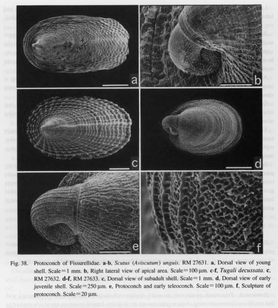
Tugali decussata (A. Adams, 1852)
(Figs. 38 c-f)
Protoconch: Protoconch paucispiral, discoidal, slightly orthostrophic. Outer surface ornamented with fine net-like sculpture consisting of ten regular-spaced spiral riblets. Lateral pouch on apical side marked by three or more oblique ribs in discordant direction to spiral sculpture. Protoconch-teleoconch boundary demarcated by thick apertural lip of protoconch.
Zeidora calceolina A. Adams, 1860
(Figs. 39 a-b)
Protoconch: Protoconch nearly symmetrical, planispiral, longitudinally elongated. Exterior sculpture mostly abraded in specimen investigated; fine granules observed at least in apical area. Protoconch-teleoconch boundary clearly demarcated, from which radiating thread-like sculpture appears near selenizone. Protoconch slightly inclined to right relative to growth axis of teleoconch.
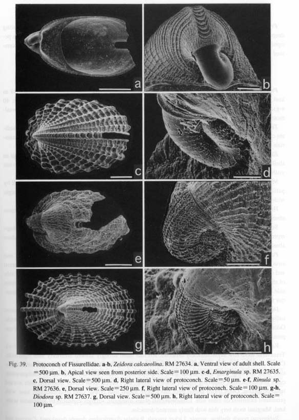
Emarginula sp.
(Figs. 39 c-d)
Protoconch: Protoconch elongated, paucispiral, almost planispiral. Lateral pouch clearly formed laterally; suture of protoconch very clear. Outer surface ornamented with fine frame-like, irregular deposits arranged axially at constant intervals. Protoconch-teleoconch boundary demarcated by strong axial ribs.
Rimula sp.
(Figs.39 e-f)
Protoconch: Protoconch paucispiral, planispiral. Outer surface ornamented by rough reticulated sculpture consisting of irregular axial and regular spiral ribs. Protoconch-teleoconch boundary clearly demarcated by distinct apertural lip of protoconch.
Diodora sp.
(Figs. 39 g-h)
Protoconch: Protoconch nearly paucispiral, planispiral. Lateral pouch weakly constricted, producing deep sutural line. Sculpture consisting of many prominent axial ribs crossed by several spiral cords on periphery. Discordant rib formed on apical side. Protoconch-teleoconch boundary unclear in specimen examined due to abrasion.
Macroschisma dilatatum (A. Adams, 1851)
(Figs. 40 a-h, 41 a-c)
Protoconch: Protoconch completely symmetrical, with posterior end strikingly projecting outward as keel (Figs. 40 b-g). Lateral pouch well-inflated, marking bilaterally deep longitudinal depression (Figs. 40 e-g). Most of surface entirely smooth. Lateral pouches striated by nearly parallel linear sculpture; dorsalmost one more distinctly prominent than others (Figs. 40 g-h).
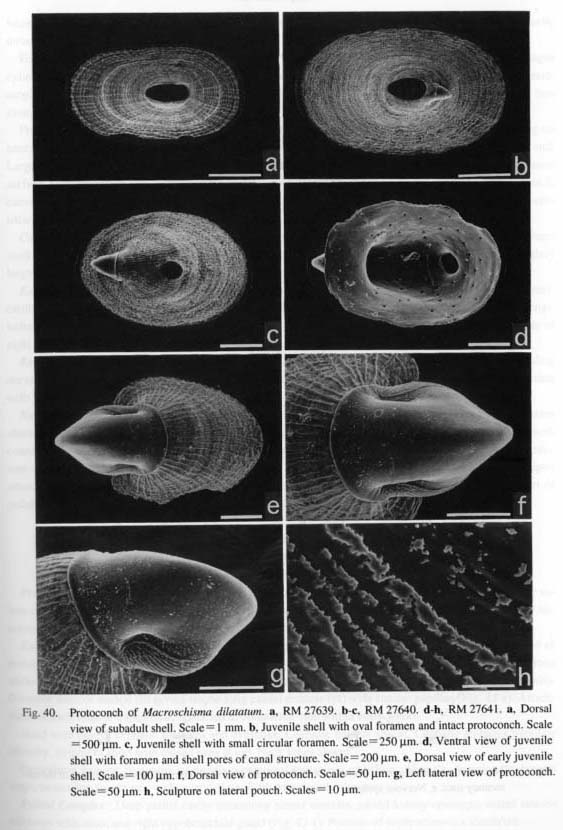
Teleoconch: At shell length of 0.6 mm, median apertural margin reflected to form foramen by immediate closure. Perforation centrally maintained by forward resorption and backward re-secretion of shell materials (Figs. 40 a-d).
External Anatomy: Shell and soft body completely limpet-shaped. Animal more than twice as large as shell in length. Because post-pericardial region wholly covered by extended epipodium, visceral hump not visible from dorsal surface.
Mantle skirt perforated by longitudinal apical hole (ah. Fig. 41 a). Mantle skirt suspended from shell by pallial retractors that mark several attachments. Mantle margin (mm) slightly reflected over shell margin with irregular microprojections.

Head with short snout (sn), non-papillate cephalic tentacles (ct), and eyestalks. Oral and cephalic lappets absent. Cephalic tentacles non-papillate. Eyes closed.
Surface of epipodium (epd) bearing tuberculate texture. Some enlarged tubercles on lateral area perhaps representing minute epipodial tentacles. Presence or absence of epipodial sense organs unclear. Operculum absent. Shell muscle (sm) horseshoe-shaped, undivided into bundles.
Pallial Complex: Pallial cavity containing paired ctenidia and osphradia, paired kidney openings, and anus. Hypobranchial glands absent. Ctenidia bipectinate, symmetrically paired in pallial cavity. Afferent and efferent ctenidial axes connected by long membranes to pallial roof along apical hole and to inner side of shell muscle, respectively. Ctenidial lamellae depressed dorsoventrally with apices projected on afferent side. Lamellae provided with bursicles and skeletal rods on efferent side. Osphradia at efferent axes of anterior free portions.
Digestive System: Composition of buccal musculature (oral, odontophoral, and radular muscles) essentially as in Scutus. Jaws paired on dorsal side of oral tube, with brush-like projections at anterior margin. Odontophore containing longer anterior and smaller posterior cartilages, connected by ventral and outer approximator muscles.
Radular sac short, posterior end deeply bifurcated. Radular formula (n+1)-(1+4)-1-(4+1)-(1+n). Transverse tooth row asymmetrically distorted, left half more strongly skewed anteriorly than right side. Central tooth plate-like, remarkably asymmetrical in both configuration and tooth morphology, without distinct shaft and cusp. Four inner lateral teeth of almost equal width, also asymmetrical in configuration; cusps non-denticulate, becoming smaller toward outside; each lateral tooth except innermost with inward basal projection. Outer (fifth) lateral teeth much more robust than others; cusp tri-denticulate; base with prominent extension on inner side, Lateromarginal plates triangular, strongly carinated by prominent acute keel. Marginal teeth very thin with finely serrated denticles.
Sublingual pouch shallow, simple. Licker smooth. Radular diverticulum deeply developed. Caecum not
found between paired odontophoral cartilages. Paired salivary glands (sg) opening into buccal cavity through longitudinal slits without ducts.
Posterior half of odontophore covered by enlarged esophageal pouches (ep) (Fig. 41 b). Mid-esophagus cylindrical. Rotation of dorsal and ventral folds gradual. Ventral folds fused in middle part of midesophagus. Inner wall covered by papillae throughout its length. Posterior esophagus (pe) opening into stomach on left side.
Proximal chamber of stomach (st) swelling to become pyriform (Fig. 41 b). Digestive glands opening on lateral area separately. Diminutive crescent-shaped gastric caecum projecting ventrally at posterior end. Large gastric tooth arising on posterior wall. Sorting area and typhlosoles well developed on ventral inner surface. Distal tapering area containing tip of protostyle. Intestine (i) extending backward beneath stomach, curving forward to produce single anterior loop (alp) (Fig. 41 b); rectum penetrating pericardium and ventricle; anus (a) opening in posterior median end of pallial cavity.
Circulatory System: Pericardium (pc) lying between apical hole and posterior margin of horse-shape shell muscle (Fig. 41 a). Heart consisting of paired auricles and median ventricle. Right auricle slightly larger than left one.
Excretory System: Kidneys paired. Right kidney spreading into posterior and anterior right sides of peri cardium. Left kidney greatly reduced, in front of left auricle. Opening of right kidney papillate, with longitudinal slit; that of left kidney minute simple pore. Right renopericardial duct extending from right side of right kidney. Left renopericardial duct not found.
Reproductive System: Gonochoristic gonad beneath digestive glands and stomach. Gonoduct ascending dorsally from right side, connecting with right renopericardial duct. Testis fine cotton-like; ovary granulate with yolk-rich eggs.
Nervous System: Circumesophageal nerve ring hypoathroid (Fig. 41 c). Labial ganglia and commissure absent. Short visceral loop arising from right and left pleural ganglia. Pedal cords (pcd) closely juxtaposed, connected with especially thick median pedal commissure (mpc). Cords on anterior side of medial commissure markedly thickened, united by about eleven thin commissures. Posterior part of cords rather abruptly attenuating. Fine thread-like pedal nerves radiating toward margin of foot. Pedal cords and most part of pedal nerves not buried in pedal musculature. Statocysts present on anterodorsal part of pedal ganglia.
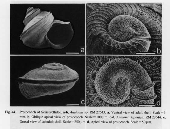
Family Scissurellidae Gray, 1847 Anatoma sp.
(Figs. 42 a-d, 43 a-h, 44 a-b)
Protoconch: Protoconch paucispiral, nearly planispiral. Weak constriction generating rather unclear suture near aperture on apical side (Fig. 44 b). Surface covered with regular net-like sculpture formed by anastomosis of fine ridges. Protoconch-teleoconch boundary demarcated by faintly thickened lip.
External Anatomy: Animal trochiform. Mantle deeply sinuate in median line (Fig. 42 a). Inner fold of mantle margin fringed by densely papillate, short microtentacles (Figs. 43 b, c). Papillae composed of long stalks and distal tuft of cilia, both of which indistinguishable from those of cephalic and epipodial tentacles. Posterior limit of mantle slit giving rise to long pallial tentacle (pt) with similar papillae (Fig. 42 a). Attachment of shell muscle trapezoidal, restricted to columellar side within body whorl.
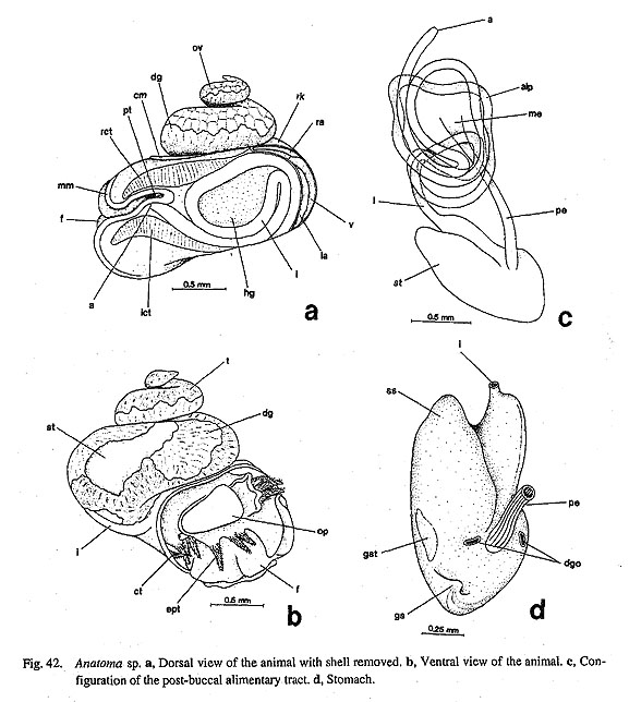
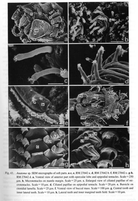
Head with pair of cephalic tentacles and eyestalks. Cephalic lappets absent. Cephalic tentacles papillate laterally. Eyestalks short with closed eyes at top. Neck lobes absent.
Epipodium bearing five pairs of papillate epipodial tentacles (ept) (Figs. 43 a, d). Anterior three pairs large, tentacular; fourth pair with extended tip. Fifth pair slightly slender.
Pallial Complex: Deep pallial cavity containing paired ctenidia, paired kidney openings, coiled intestinal loops with anus, and right hypobranchial gland (Fig. 42 a). Position of osphradium not identified.
Ctenidia (rct, lct) slightly asymmetrical, composed of about thirty monopectinate lamellae. Lamellae covered by cilia zoned laterally. Bursicles (br) prominently developed near efferent axis (Fig. 43 e). Opening of lumen slit-like, continued to lateral cilia. Few tufts of cilia on exterior part of bursicle. Hypobranchial gland (hg) lying within pallial loop of intestine, restricted to right side relative to rectum, consisting of tall cells containing spherules.
Digestive System: Oral tube surrounded by buccal sphincter and constrictor (bs, bct), fixed by mandibular protractors and mandibular retractors. Muscles of odontophore comprising lateral protractors (lp), anterior levators (al), posterior depressors (pd), and ventral protractors (ovp, ivp) (Fig. 43 f). Posterior levators absent. Dorsal posterior part united by postdorsal (pdt) and dorsal buccal tensors (dbt).
Jaws consisting of paired elements attaching to oral tube posteriorly but projecting into mouth anteriorly.
Anterior margins indented with scaly projections.
Radular sac form single vertical loop behind buccal mass. Posterior end clearly bifurcated. Radular formula n-(1+4)-1-(4+1)-n (Figs. 43 g, h). Transverse radular row symmetrical. Central tooth broadest of all radular teeth; base broad, extending over innermost lateral teeth; shaft tapering distally, cusp indented by one thick median and seven thinner lateral denticles. Inner lateral teeth progressively reducing in width outwardly; shafts strongly bent at base; cusps triangular-shaped, sharply denticulate; inner three laterals of common shape with five or six. denticles, but slender outermost one with only three denticles. Outer (fifth) laterals with broad base, articulating with bases of inner laterals; cusp sharply hooked along outer margin, finely serrated by small denticles, Lateromarginal plates absent. Marginal teeth slender, thread-like, with less than six denticles on cusps.
Subradular membrane connected to lateral and median protractors (lpr, mpr) and retractors (rsr). Thin tensor muscle of radular sac (trs) running from dorsal part of sublingual pouch to ventral side of radular sac posterior to buccal mass (Fig. 43 f). Postmedian retractors of radular sac absent.
Odontophore containing elongated anterior and small posterior cartilages connected by ventral and outer approximator muscles.
Sublingual pouch shallow, simple. Radular diverticulum deeply developed. Salivary glands attached to buccal cavity dorsally without ducts. Anterior esophagus inflated posterior to buccal mass. Mid-esophagus (me) not very long; counterclockwise rotation of dorsal and ventral folds apparent. Inner wall covered with papillae except in ciliated dorsal food channel. Posterior esophagus (pe) extending along columella on right side, corrugated by several longitudinal furrows internally.
Stomach (st) large for body size, visible from dorsal surface of visceral hump (Fig. 42 b). Posterior apex slightly curving to form short gastric caecum (Fig. 42 d). Paired folds and median groove visible through outside of caecum. Digestive glands (dg) opening on either side of end of posterior esophagus. Distal portion of stomach distinctly divided into isolated style sac region (ss) and preintestinal extension. Tooth of gastric shield (gst) lying at base of style sac region; paired typhlosoles running through extended part of preintestinal region.
Intestine (i) running complicated course through both visceral and pallial regions (Fig. 42 c). Anterior loop (alp) formed beyond mid-esophagus. After penetrating into pericardium and ventricle, pallial section forming large loop anteriorly. Loop in pallial cavity recurving once, finally terminating on left side of mantle slit.
Circulatory System: Pericardium elongated along posterior limit of pallial cavity. Heart consisting of right auricle (ra) lying at right posterior side, left auricle (la) on left anterior side, and median ventricle (Fig. 42 a).
Excretory System: Kidneys asymmetrically paired. Right kidney (rk) lying on right side of right auricle along columella; presence indicated by light brown color in fixed material. Left kidney (lk) on left side of pericardium; lumen lacking distinct papillae.
Reproductive System: Gonad (ov, t) lying on dorsal side of digestive glands in spiral part of visceral hump (Figs. 42 a, b). Sexes apparently separated, distinguished by texture of granulation of gonadal tissue. Pathway of gonoduct to pallial cavity not determined. Copulatory organ absent.
Nervous System: Circumesophageal nerve ring hypoathroid. Pedal ganglia nearly fused in midline. Pedal cords buried in pedal musculature without median connection (unlike in Scutus and Macroschisma). Labial ganglia and commissure not found. Visceral loop originating directly from pleural ganglia. Statocysts on anterior sides of pedal ganglia.
Anatoma japonica (A. Adams, 1862)
(Figs. 44 c-d)
Protoconch: Protoconch paucispiral, almost planispiral. Suture unclear, represented only by spiral depression. Outer ornament consisting of irregularly arranged granules. Protoconch-teleoconch boundary margined by delicate axial line.
Sinezona sp.
(Figs. 45 a-c)
Protoconch: Protoconch spirally coiled, hyperstrophic. Terminal part of protoconch oriented more dorsally over initial part at distinct angle. Surface very strongly sculptured. On dorsal side, at least fifteen tall varices arranged in parallel to apertural margin. Characteristically, most centrally situated rib with quite different angle to remaining ribs. This rib much longer than others extended from dorsal to ventral centers. Ribs formed as aggregations of tall spicular prisms.
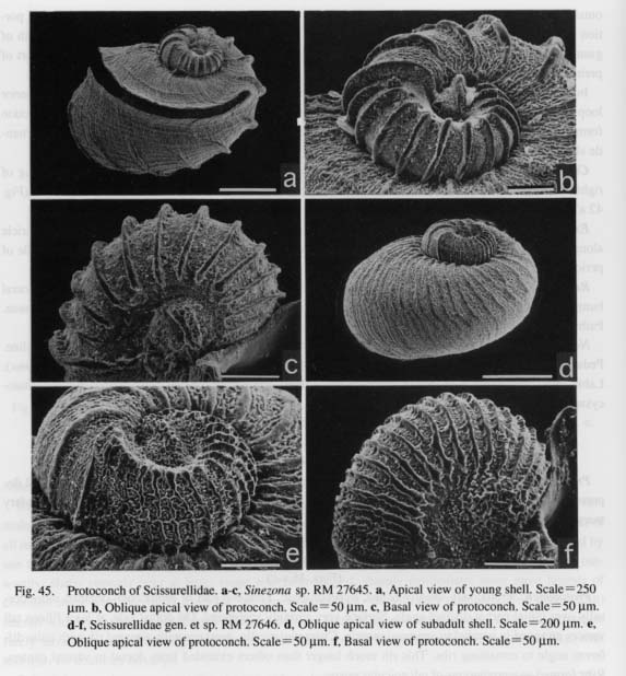
Scissurellidae gen. et sp.
(Figs. 45 d-f)
Protoconch: Protoconch paucispiral, with coiling direction nearly planispiral but slightly hyperstrophic. Surface covered with dense series of axial ribs transversely connected with weak spiral ridges. Interspaces between axial ribs entirely filled with fine granules. In central part of protoconch, aggregation of granules forming distinct ridge-like structure on both apical and basal sides. Protoconch-teleoconch boundary marked by thicken axial lip.
Family Turbinidae Rafinesque, 1815
Turbo (Marmarostoma) stenogyrum (Fischer, 1873)
(Figs. 46 a-b, 47 a-f, 48 a-b)
External Anatomy: Mantle margin (mm) lacking microtentacles. Shell muscle consisting of single undi-vided columellar muscle (cm).
Head with snout (sn), cephalic lappets (clp), papillate cephalic tentacles (ct), and eyestalks (es). Outer lip of mouth forming thick oral disk. Eyes (e) open, plugged with vitreous body at tips of eyestalks. Inhalant (left) and exhalant (right) neck lobes present in neck region.
Foot (f) with four pairs of long papillate epipodial tentacles, each with knobby epipodial sense organ at base. Head-foot folding anteroposteriorly when retracted. Calcareous operculum attached to opercular lobe of epipodium.
Pallial Complex: Pallial cavity extending more than half of body whorl, including left ctenidium, left osphradium, paired kidney openings, anus, and paired hypobranchial glands (Figs. 46 a, b).

Ctenidium (c) extending almost entire length of pallial cavity, attached to left wall of pallial cavity by efferent ctenidial membrane and to pallial roof by afferent ctenidial membrane (acm). Left dorsal area of pallial cavity divided by afferent ctenidial membrane, generating isolated pocket space dorsally. Ventral ctenidial lamellae more enlarged and projecting to right side than dorsal lamellae. Efferent side of lamellae provided with series of bursicles (br, Fig. 47 a) and skeletal rods (sr) internally (Fig. 47 c).
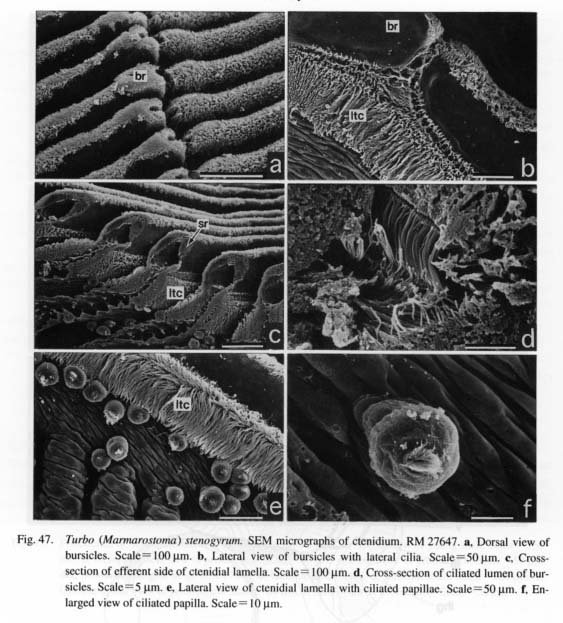
Bursicles prominent, arrayed longitudinally along efferent ctenidial axis. Presence of bursicle marked by dorsally expanded knob and deep ciliated groove (Figs. 47 a, b). Inside of bursicle with pocket lumen, heavily ciliated with long (7-8 µm) simple cilia (Fig. 47 d). Exit of pocket lumen visible from dorsal side as vertical slit. On sides of ctenidial filament, ciliated micropapillae (ca. 20 µm in diameter) occurring be-neath strip of lateral cilia (Figs. 47 e, f).
Osphradium covering free position of efferent ctenidial axis. Paired hypobranchial glands on pallial roof on either side of rectum. Right gland (rhg) much smaller than left gland. Left gland (lhg) intersected by transverse pallial vessel dorsally.
Digestive System: Oral tube surrounded by buccal sphincter (bs) and constrictor (bct) with mandibular protractors (mp) and retractors (mr). Movement of odontophore controlled by lateral protractors (lp), inner and outer ventral protractors (ovp, ivp), anterior levators (al), and posterior depressors (pd) (Figs. 48 a, b). Posterior levators absent. Right and left sides tightly held by postdorsal buccal tensor (pdt) and dorsal buccal tensors (dbt).

Jaws (j) divided into symmetrical pairs on dorsal side of oral tube. Anterior edge fimbriate, projecting into oral cavity.
Radular sac forming coiled loops behind buccal mass, posterior end clearly bifurcated. Radular formula n-5-l-5-n. Radular tooth row symmetrical. Central tooth subrectangular, plate-like, with indistinct cusp. Inner four lateral teeth slender with reduced cusps and outward projection at bases. Outermost laterals spatulate, almost straight without basal projection. Bases of laterals overlapping from inner to outer teeth. Protolateromarginal plates not well-developed. Inner marginals prominently elongated, but outer teeth gradually diminishing in size.
Manipulation of radula controlled by median and lateral protractors of subradular membrane (mpr, lpr), retractors of subradular membrane (rsr), retractors of radular sac (rrs), and postmedian retractors of radular sac (prs) (Fig. 48 b). Radular sac connected to sublingual pouch ventrally by tensor muscle (trs) which arises from space between paired postmedian retractors (prs).
Buccal mass containing two pairs of cartilages. Anterior cartilages elongated, connected to ventral ap
proximator. Posterior cartilages small, extending laterally. Both cartilages united longitudinally by outer approximators.
Sublingual pouch shallow without glandular outgrowth. Radular diverticulum present, Buccal cavity overlain directly by salivary glands without ducts, followed by large esophageal pouches overlapping pos-terior part of buccal mass. Mid-esophagus long, gradually reduced in width posteriorly. Dorsal and ventral folds clearly formed within it. Inner wall covered with papillate esophageal glands except in dorsal food channel. Posterior esophagus short.
Stomach bent, divided into small proximal and large distal sacs. Posterior apex of stomach with promi-nent spiral caecum (gc) visible from surface of visceral hump (Fig. 46 b). Distal portion including openings of digestive glands, toothed gastric shield, paired typhlosoles, and intestinal groove. Corrugated pattern of sorting area visible from exterior. Intestine coiling complexly with single anterior loop. Rectum penetrating ventricle, running along pallial roof, opening anteriorly.
Circulatory System: Pericardium within visceral hump along posterior limit of pallial cavity (Figs. 46 a, b) containing paired auricles and median ventricle. Right auricle (ra) narrowly elongated; left auricle (la) well inflated. Aortae bifurcated within pericardium without forming bulbous aorta.
Anterior aorta running along efferent ctenidial vessel on pallial wall, entering head region. Posterior aorta passing within digestive glands.
Blood from right kidney draining posteriorly into left afferent renal vein (arv), anteriorly into transverse pallial vessel (tpv) penetrating left hypobranchial gland, reaching afferent axis of ctenidium. Afferent cteni-dial vessel (acv) supplying blood to afferent side of ctenidial lamellae, also giving rise to branch into ante-rior pallial vessel (apv) along margin of afferent ctenidial membrane (acm). Blood from ctenidial lamellae and mantle skirt entering efferent ctenidial vessel (ecv), leading to ventricle. Efferent renal vessel also entering ventricle from left posterior end of left kidney.
Excretory System: Excretory system consisting of right and left kidneys (Figs. 46 a, b). Right kidney (rk) within visceral hump, also extending to right side of anterior visceral region over mid-esophagus; inner wall lamellate. Right kidney opening short papilla. Left kidney (lk) occurring dorsally within pallial cavity, inner wall papillate. Left kidney opening represented by longitudinal slit.
Reproductive System: Gonad (g) on dorsal side of digestive glands (Figs. 46 a, b), when ripe, reaching posterior margin of right kidney. Gonoducts connected to right kidney, but relationship with renopericardial duct not identified. Sexes distinguished by external appearance. Ovary clearly granular; testis exhibiting unclear granulation.
Nervous System: Circumesophageal nerve ring hypoathroid. Cerebral ganglia at bases of cephalic tentacles; pleural and pedal ganglia on posteroventral sides of buccal mass. Cerebral commissure overlapping frontal margin of buccal cavity. Labial ganglia and labial commissure absent. Buccal ganglia connected to ventral extension of cerebral ganglia.
Paired pleural ganglia sending nerves to outer sides of body musculature including columellar muscle. In left anterior pallial wall, nerves from supraesophageal to osphradioctenidial ganglia united by distinct nerves of zeugoneury from anterior pallial nerve. Visceral loop originating from right and left pleural ganglia, elongated along pallial cavity wall. Differentiation of supraesophageal and subesophageal ganglia not very apparent. Pedal cords well developed with commissures. Statocysts on anterior dorsal sides of pedal ganglia.
|