Family Patellidae Rafinesque, 1815
Patella (Penepatella) flexuosa Quoy and Gaimard, 1834
(Figs. 1 a-b, 2 a-d)
External Anatomy: Mantle margin (mm) provided with numerous short circumpallial tentacles (pt) (Figs. 1 a, b). Shell muscle (sm) horseshoe-shaped, divided into about sixteen bundles by blood vessels from visceral hemocoel to circumpallial vessel (Fig. 1 a). Pallial retractor muscle connecting shell muscle anteriorly.
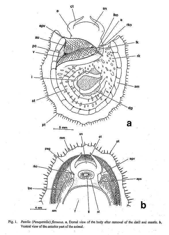
Head with short rounded snout (sn) and pair of smooth cephalic tentacles (ct) without papillae (Figs. 1 a, b). Margin of outer lip (ol) lacking microtentacles and oral lappets (Fig. 1 b). Eyes (e) open, longitudinally invaginated, black pigmented within bases of cephalic tentacles.
Epipodial region lacking tentacles and sensory projections. Operculum absent. Two distinct sensory streaks present on lateral walls of shell muscle (Fig. 1 b). Lateral pallial streaks (Ips) extending posteriorly on sides of foot. Anterior pallial streaks (aps) much shorter than lateral ones, attached to anterior ends of shell muscle; inner ends turning slightly inward to pallial cavity.
Pallial Complex: Pallial cavity shallow, restricted to narrow area of nuchal region, containing "secondary" pallial gills, paired kidney openings, anus, and paired osphradia. Ctenidium and hypobranchial gland absent, Pallial gills (pag) arising from circumpallial vessel by evagination (Fig. 1 b). Respective leaflets triangular, feather-like, with tips directed to outside. Outer edge of leaflets thicker and more stiffen than inner margin. Floor of pallial cavity with pair of osphradia on inner sides of anterior shell muscle ends. Tuberculate wart-organ well developed in outer part of osphradia.
Digestive System: Buccal sphincter and constrictor not clearly developed. Very thin pairs of mandibular protractors and retractors inserted on jaw plate. Movement of inner lips (il) controlled by transverse labial muscles. Extrinsic muscles of odontophore comprising dorsal protractors, lateral protractors, ventral protractors, and anterior levators. Depressors absent. Posterior part of buccal mass lacking tensor muscle.
Jaw simple single plate, consisting of larger posterior and smaller anterior wings (aw, pw, Fig. 2 a); their overlapping area thickened to form smooth anterior band (ab). Posterior wings affixed to odontophore by muscle attachments.
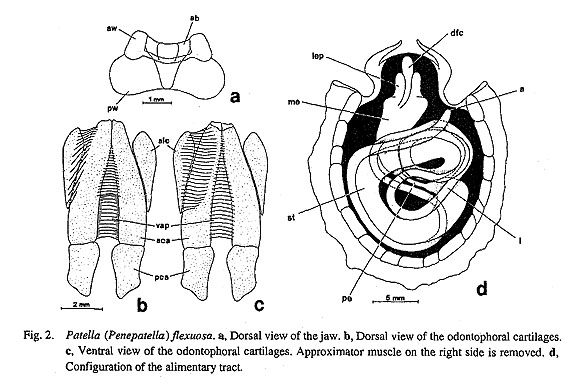
Radular sac long, turning in a few loops. Posterior end of radula sac simple. Radular formula 3−(1 +2)− 1−(2+1)−3 (see Sasaki et al., 1994 for SEM micrographs). Radular row symmetrical. Central tooth extremely thin, represented only by chitinous ridge between innermost lateral teeth, without any trace of mineralization. Lateral teeth consisting of two pairs of slender inner teeth with simple cusps and one greatly enlarged pair of outer teeth each with four denticles. Inner two marginals vertically aligned. Outermost marginals broader, situated more posteriorly than inner teeth. Basal plates complexly sculptured by thick ridges that continue to bases of teeth.
Muscles of subradular membrane and radular sac exhibiting characteristically complicated configuration. Subradular membrane controlled by six muscles: (1) Median protractors of subradular membrane, with markedly asymmetrical arrangement. Anteriorly right and left muscle strands running backward between cartilages along radular sac. In posterior area of buccal mass, right muscle bending sharply toward right, penetrating membrane separating cephalic and visceral sinuses, finally merging into right shell muscle. Left
muscle running straight posteriorly along radular sac. (2) Lateral protractors of subradular membrane, originating at anterior end of subradular membrane, inserting onto posterior cartilages (pca). (3) Retractors of subradular membrane composed of two layers (dorsal and ventral) on ventral side. (4) Retractors of radular sac, connecting to ventral side of radular sac and posterior end of radular cartilages. (5) Tensor of radular sac, originating from ventral surface of radular sac far behind buccal mass, inserting in dorsal part of retractors of subradular membrane. (6) Median retractor of radular sac (mrs), running along left strand of median protractor (mpr).
Odontophoral cartilages mainly consisting of three pairs (Figs. 2 b, c). Anterior cartilages (aca) longitudinally elongated, slightly curved outward. Posterior cartilages (pac) half as long as anterior cartilages, subequal in width. Anterolateral cartilages (ale) contacting but not overlapping with anterior cartilages. Ventrolateral and dorsal posterior cartilages (vlc, dca) greatly reduced. Labial cartilages (as in Cellana, below) absent.
Anterior cartilages connecting to two-layered ventral approximator (vap). Insertion of dorsal layer restricted to anterior half of anterior cartilages, while that of ventral layer extending along length of anterior cartilages (Figs. 2 b, c).
Sublingual pouch lacking outgrowths of sublingual gland. Licker sharply lamellate. Radular diverticulum wide and deep between anterior esophagus and radular sac. Paired salivary glands completely fused as single glandular mass behind buccal mass. Two pairs of salivary ducts extending from anterior margin of salivary glands. Inner ducts running along anterior esophagus, emptying at posterior end of buccal cavity. Outer ducts running along outer sides of buccal mass, opening into lateral walls of buccal cavity.
Anterior esophagus divided into median dorsal food channel (dfc) and lateral esophageal pouches (lep) over buccal mass by ventral and dorsal folds (Fig. 1 d). Lateral pouches extended to form well-expanded mid-esophagus (me). Mid-esophagus gradually curving to right, reducing its width toward central part of visceral mass, then sharply constricting into narrow posterior esophagus. Mid-esophagus densely septated internally by transverse septum-like evagination of glandular epithelium. Posterior esophagus (pe) striated internally by longitudinal ridges whose pattern is visible from outside.
Stomach (st) curving in C-shape along posterior and left shell muscle (Fig. 1 d). Digestive glands opening into initial chamber of stomach through paired ducts. Stomach totally lacking gastric caecum, sorting area, tooth of gastric shield. Very thin typhlosoles running along length of stomach. Intestine (i) with four loops, two of them anteriorly shifted. Rectum running along pericardium without penetration. Anus (a) opening at right posterior comer of pallial cavity.
Circulatory System: Heart containing single auricle, ventricle, and muscular bulbous aorta (Fig. 1 a). Auricle (au) receiving blood only from left side via efferent pallial vessel (epv). Anterior margin of auricle fixed to anterior wall of pericardium, its left wall more prominently expanded than right wall. Ventricle (v) obliquely posterior to auricle, connected to posterior wall of pericardium by connective tissues. Bulbous aorta completely ventral to ventricle. Venous blood flowing from kidneys to circumpallial vessel through afferent pallial vessels penetrating shell muscle. Circumpallial vessel extending to anterior pallial cavity as complete circle, provided with secondary pallial gills (pag) (Fig. 1 b). Efferent pallial vessel (epv) entering auricle only from left side.
Excretory System: Excretory organs consisting of right and left kidneys (Fig. 1 a). Left kidney (lk) much smaller than right, restricted to small area between rectum and pericardium. Right kidney (rk) very large, entirely enclosing digestive glands both dorsally and ventrally. Both kidneys with papillate opening (rko, lko) on either side of anus. Papilla of right opening more prominently extended than that of left opening. Right opening vertically slit-like; left simple.
Reproductive System: Gonad developing below digestive system, emerging dorsally on left side when gravid. Gonoduct arising from left side, passing below rectum, discharging into right kidney independent of left renopericardial duct. Right kidney opening functioning as urinogenital opening. Sexes separate. Testis creamy in color; ovary reddish brown.
Nervous System: Circumesophageal nerve ring hypoathroid. Cerebral ganglia at bases of cephalic tentacles. Pleural and pedal ganglia below posterior end of buccal mass, connected to cerebral ganglia by long connectives. Oral region containing labial ganglia with distinct labial commissure. Buccal ganglia connected to labial ganglia through labiobuccal connectives. Supraesophageal, subesophageal, and visceral ganglia fused as tightly twisted visceral loop on right side. Supra-and subesophageal parts of visceral loop supplying nerves to osphradial ganglia. Zeugoneury absent. Circumpallial nerve forming complete circle. Pedal cords with many incomplete pedal commissures. Statocysts on outer ventral sides of pedal ganglia.
Family Nacellidae Thiele, 1891
Cellana toreuma (Reeve, 1854)
(Figs. 3 a-c, 4 a-b, 5 a-d, 6,7 a-b, 8 a-b, 9 a-b, 10 a-h, 11 a-h,)
Protoconch: Protoconch pear-shaped with well-inflated apex, entirely symmetrical along longitudinal axis (Figs. 3 a-c). Adapertural part elongated, somewhat constricted. Exterior surface with reticulate sculpture creating pit-like appearance between ridges. On dorsal side, network ridges anastomosing to exhibit denser sculpture. Eight longitudinal streaks (one dorsal and ventral in midline, three on each side), arising and radiating from posteroventral tip of protoconch.

External Anatomy: Animal limpet-shaped without visceral coiling. Mantle margin (mm) fringed with series of circumpallial tentacles (Fig. 4 a, pt) consisting of regularly alternating longer and shorter ones. Tentacles pigmented by gray circular bands (Fig. 4 b). Under SEM observation, clusters of cilia scattered on surface of tentacles (Figs. 10 a, b). Attachment of shell muscle (sm) horseshoe-shaped, constricted into about sixteen bundles penetrated by vessels (Fig. 4 a). Pallial muscle connecting right and left ends of shell muscle beyond head.
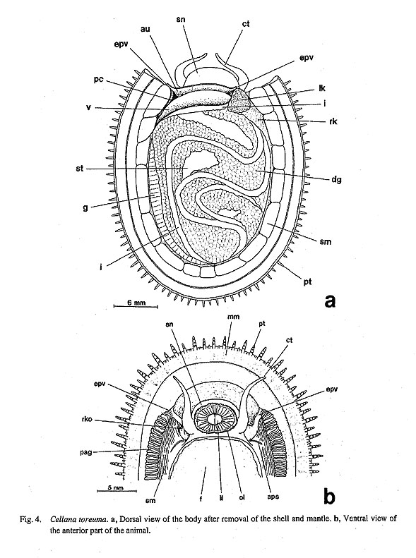
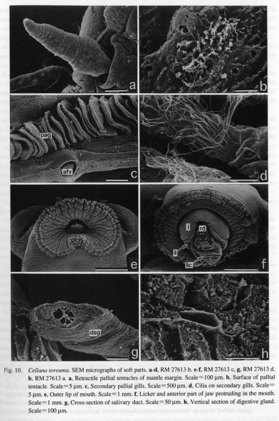
Head with short snout (sn) and pair of non-papillate cephalic tentacles (ct). Snout always directed ventrally, fitting mouth to substratum. Outer lip (ol) of mouth fringed with minute tentacles (Fig. 10 e). Inner lips (il) arising from inner lateral wall of oral tube, with only median parts exposed. Oral lappets absent. Eyes open, simply invaginated cups, with inner basal portions black-pigmented; presence clearly indicated externally by short slits.
Tentacles, sense organs, and operculum absent. Only one pair of sensory streaks in pallial groove; anterior pallial streaks (aps) running along both ends of shell muscle, extending slightly backward in pallial groove. Lateral pallial streaks (as in Patella, above) absent.
Pallial Complex: Pallial cavity shallow, restricted to small space in nuchal region, containing "secondary" pallial gills, anus, paired kidney openings, and paired osphradia. Ctenidium and hypobranchial gland absent.
Pallial gills (pag) developing from circumpallial vessel (Fig. 10 c), with cilia arising irregularly from its surface (Fig. 10 d). Gills very characteristically absent in front of head where circumpallial vessel interrupted (Fig. 4 b).
Paired osphradia symmetrically situated on floor of pallial cavity near anterior end of shell muscle; right and left osphradia of subequal size. Surface of wart-organ (wo) smooth, lacking ciliated zone, with spongy structure in cross-section (Figs. 9 e, f, SEM observation).
Digestive System: Oral tube not surrounded by circular buccal sphincter and constrictor, but instead covered by jaw. Inner lateral walls of oral cavity giving rise to thickened inner lips which are fixed by transverse labial muscles (tlm, Fig. 5 c).
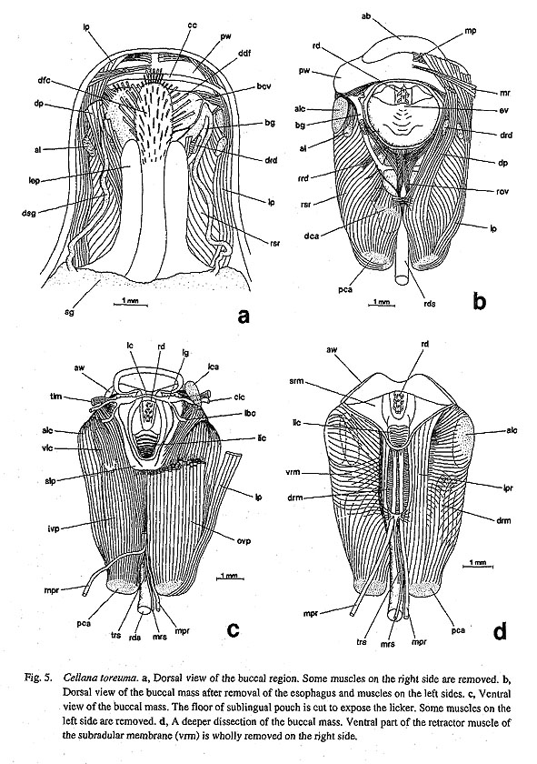
Odontophore connected to wall of body cavity by dorsal, lateral, and ventral protractors, and anterior levators (Figs. 5 a-d, 6): (1) Dorsal protractors (dp) occupying most dorsal part of buccal mass, running obliquely along anterior esophagus, inserting on posterior cartilages. (2) Lateral protractors (lp) running along outer sides of odontophore over retractors of subradular membrane (rsr). (3) Ventral protractors (vp) distinctly divided into two layers. Outer ventral protractors (ovp) as most ventral part of musculature, connected to body cavity floor beneath sublingual pouch. Inner ventral protractors (ivp) widely enveloping ventral side of odontophore, attached to posterior end of sublingual pouch, margin or inner surface of ante
rior and posterior wings of jaw, ventral side of oral tube, and part of retractors of subradular membrane. (4) Anterior levators (al) inserting on anterolateral sides of odontophoral cartilages, running along anterior end of cartilages below labiobuccal connective, rising dorsally toward origin on roof of body cavity. Depressors and tensors absent in posterior part.

Jaw forming single plate composed of smaller anterior and larger posterior wings (aw, pw) (Fig. 7 b). Anterior margin thickened into smooth anterior band (ab). Most parts of jaw covering dorsal side of oral tube. Posterior wings firmly fixed to odontophore by muscle attachment.

Radular sac very long, forming more than three loops. Posterior end of radular sac not bifurcated. Radular formula 1?-2-1-2-1?. Inner lateral teeth thin with acute cusp and central ridge. Cusp of outer lateral teeth divided into two lateral parts; acutely pointed inner cusp with median ridge; blunt outer cusp smoothly continued to outer margin of shaft, forming excavated longitudinal cleft. Marginal teeth represented by thin plates adhered to radular membrane. Basal plates sharply sculptured.
Subradular membrane fixed by thick retractors and two relatively thin pairs of odontophoral protractors: (1) Retractors of subradular membrane (rsr) extending from lateral margin of subradular membrane, entirely enclosing cartilages, ventrally divided into two layers (Fig. 5 d): dorsal layer (drm) between two pairs of subradular membrane protractors and ventral approximator of odontophoral cartilages; ventral layer (vrm) fused between inner ventral protractors of odontophore and two pairs of subradular membrane protractors. (2) Median protractors of subradular membrane (mpr) extending backward along midline; left protractor running straight, but right one more or less sharply bending left, ventrally penetrating buccal membrane, ultimately attaching to right shell muscle (Figs. 5 c, d). (3) Lateral protractors of subradular membrane (Ipr) lying laterally to median protractors, inserting onto posterior cartilages (pea) (Fig. 5 d).
Ventral side of radular sac inserted by (1) paired retractors of radular sac (rrs) (Fig. 6), (2) unpaired median retractor of radular sac (mrs), and (3) unpaired ventral tensor of radular sac (trs) (Figs. 5 c, d).
Odontophore containing five pairs of cartilages: anterior (aca), posterior (pca), anterolateral (alc), ventrolateral (vlc), and posterodorsal cartilages (dca) (Fig. 7 a). In addition, pair of labial cartilages (Ica) on ventral floor of buccal region (Fig. 6). Odontophoral part of cartilages interconnected by sheet-like muscles to each other to form firm core of buccal mass. Among them, anterior cartilages especially tied by welldeveloped, two-layered ventral approximator muscle (vap).
Anterior part of buccal cavity giving rise to ventral branch of deep sublingual pouch (slp) without outgrowths (Fig. 5 c). Licker (lic) provided with 9-10 transverse lamellae (Figs. 5 d, 10 f). Radular diverticulum narrow, longitudinally elongated below esophageal valve. Salivary glands (sg) pouch-like, widely extended over post-buccal region (Fig. 5 a). Paired long salivary ducts (dsg) arising from outer anterior part of glands, discharging into buccal cavity bilaterally (Figs. 5 a, 10 g).
Inner portion of anterior esophagus distinctly partitioned into dorsal food channel (dfc) and lateral pouches (lep) by inner ventral folds (vf) and outer dorsal folds (df) (Figs. 8 a, b). Mid-esophagus (me) bending toward left in visceral region. Boundary between anterior and mid-esophagi clearly delimited by internal structural difference (Fig. 8 b). Dorsal and ventral folds of mid-esophagus prominently rotated counterclockwise. Right and left dorsal folds fused into single fold posteriorly; both ventral folds fused at beginning of mid-esophagus. Mid-esophagus ending with distinct constriction. Posterior esophagus (pe) internally corrugated by longitudinal grooves.

Stomach (st) consisting of smaller proximal part of sigmoidal loop and larger C-shaped distal part (Fig. 8 a). Only initial chamber of stomach with small openings of ducts from digestive glands. Sorting area indistinct; gastric caecum and tooth of gastric shield totally absent. Intestinal groove between typhlosoles very shallow. Intestine (i) consisting of more than ten loops, coiling complexly within digestive glands. Rectum not penetrating ventricle or pericardium. Anus (a) opening at right anterior corner of pallial cavity.
Circulatory System: Heart composed of auricle, ventricle, and muscular bulbous aorta (Fig. 4 a). Very
characteristically, auricle (au) receiving efferent pallial vessels from both right and left sides, plus capillary vessels from pallial cavity roof (Figs. 4 a, b). Left posterior side of auricle with narrowly expanded space, rendering its outline asymmetrical. Ventricle (v) muscular compared to auricle. Inner wall of ventricle wholly covered with granules (Fig. 11 a). Wall of auricle fixed by connective tissue to wall of pericardium (pc) on right side. Bulbous aorta obliquely elongated, ventrally connected to ventricle through very long narrow slit, leading to anterior aorta on right anterior side and to posterior aorta on left posterior side.

Anterior aorta running ventrally into buccal region below salivary glands. Behind buccal mass, anterior aorta entering radular artery which encloses whole radular sac and extends back to visceral region. Anterior aorta further extending anteriorly as buccal artery between right and left cartilages, expanding to form buccal sinus (bsn) in center (Fig. 6), separated from surrounding cephalic sinus by buccal membrane but partly continuous through lacunae.
Posterior aorta bifurcating beyond pericardium. Ventral branch supplying blood to gonad; dorsal branch supplying digestive system. Branches divaricating complexly within visceral mass, associated with right kidney surrounding visceral mass circularly both dorsally and ventrally. Venous blood vessels from visceral sinus passing though shell muscle toward circumpallial vessel, finally entering auricle through paired efferent pallial vessels.
Excretory System: Paired asymmetrical kidneys on either side of terminal part of intestine (Figs. 4 a, 11 b). Both kidneys lying on right side of pericardium. Left kidney (lk) extremely small relative to right, its inner wall covered densely with vermiform projections (Fig. 11 c). Opening of left kidney papillate with longitudinal slit (Fig. 4 b). Right kidney (rk) extending widely along shell muscle, surrounding digestive glands and stomach, its lumen spacious and partitioned by membranous structure (Fig. 11 d). Opening of right kidney (rko) forming simple pore. Left renopericardial duct opening as horizontally elongated slit at base of right posterior wall of pericardium. Right renopericardial duct opening into right wall of pericardium through minute pore.
Reproductive system: Reproductive organ consisting of very simple gonad and gonoduct. Gonad (g) situated within ventral side of visceral mass; left side extending to dorsal surface as it becomes gravid and increases its volume (Fig. 4 a). Gonoduct connected to right kidney independent of renopericardial duct. Because of its thinness, gonoduct often difficult to trace in macroscopic observation in non-breeding condition, but packed with gametes in gravid phase.
Sexes separate; consecutive hermaphroditism not observed. Male and female identified by texture of gonadal contents: granular in female (ovary), cotton-like in male (testis); this difference reflecting morphology of clusters of gametes within gonad. Spermatozoa with long head and thread-like tail (Fig. 11 g). Ova covered by thick layer of yolk (Fig. 11 h). Gametes discharged through right kidney; fertilization free in sea water.
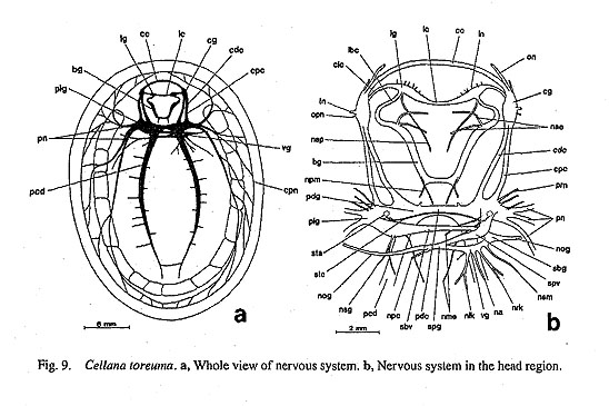
Nervous system: Circumesophageal nerve ring hypoathroid (Figs. 9 a, b). Cerebral ganglia (cg) at bases of cephalic tentacles, interconnected to cerebral commissure (cc) running in front of posterior wings of jaw. Pleural and pedal ganglia (plg, pdg) juxtaposed along posterior margin of buccal mass, each connected to cerebral ganglion by long connective (cdc, cpc). Both lying not in visceral region, but within cephalic sinus separated from visceral sinus by membrane.
Cerebral ganglia innervating laterally cephalic tentacles via tentacular nerves, eyes via optic nerves, and anteriorly to oral region of snout. Inner sides of ganglia also providing thin but distinct nerves to ventral side of buccal cavity. Labial ganglia (Ig) supplying nerves anteriorly to labial region through about 7 labial nerves and posteriorly to sublingual pouch and its vicinity (Figs. 5 c, 9 b), connected by labial commissure (Ic). Elongated buccal ganglia (bg) between esophagus and radular diverticulum, providing nerves to buccal cavity roof, dorsal food channel, and muscles of buccal mass. Buccal and labial ganglia connected through labiobuccal connectives (Ibc) running between posterior wings of jaw and lateral protractor muscles of odontophore.
Very tight visceral loop formed by fusion of visceral (vg), supraesophageal (spg), and subesophageal (sbg) ganglia just behind buccal mass on right side. Loop prominently slanted against horizontal plane of body with subesophageal side placed more ventrally. Ganglia of loop innervating kidneys (nrk, nlk), anus (na), digestive glands and tracts, and pericardium (npc). Thin nerve particularly extending to shell muscle from subesophageal side (nsm, Fig. 9 b). Connectives with osphradial ganglia also arising from sides of visceral loop.
Pleural ganglia close to lateral body walls, giving off several thin parietal nerves (pm) and thicker pallial nerves (pn). Latter nerves dividing into anterior and posterior branches on both right and left sides, penetrating shell muscles, extending into thin mantle, reaching cord of circumpallial nerve (cpn). Fine branches from pallial nerves forming complex nerve plexus by anastomosing in mantle margin. Zeugoneury absent on both sides.
Pedal ganglia not very concentrated, emitting thick pedal cords to pedal musculature. Statocysts (sta) located lateral to pedal but ventral to pleural ganglia, united by very thin commissure (stc) (Fig. 9 b).
Family Lepetidae Dall, 1869
Limalepeta lima (Dall, 1918)
(Figs. 12 a-b, 13 a-b, 14 a-c)
External Anatomy: Circumpallial tentacles of mantle margin not visible in fixed specimen; living specimens not observed. Shell muscle (sm) horseshoe-shaped with pallial retractor muscle, divided into bundles by blood vessels. Head with short snout, and non-papillate cephalic tentacles (ct) without eyestalks. Outer lip (ol) of mouth surrounded by oral lappets (olp) (Fig. 12 b). Eyes of simple invaginated type within bases of cephalic tentacles. Foot lacking lateral epipodial tentacles and sense organs, and operculum. Anterior and lateral pallial streaks also absent.
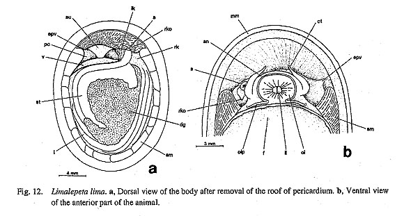
Pallial Complex: Pallial cavity shallow, containing only anus and paired kidney openings on posterior wall (Fig. 12 a). No true ctenidia or "secondary" gills. Osphradia and osphradial ganglia absent. Hypobranchial gland absent.
Digestive System: Movement of inner lips controlled by transverse labial muscles (tlm) running below
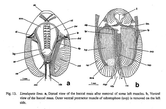
anterior wings of jaw toward origin of lateral wall of snout. Muscles of odontophore consisting of following muscles (Figs. 13 a, b): (1) Dorsal protractors (dp) running from end of posterior cartilages, passing between anterior levators and dilators of radular diverticulum (drd), attaching to margin of posterior wings of jaw. Behind buccal cavity, right and left muscles connected by thin transverse muscle tissues over retractors of esophageal valve (rov). (2) Lateral protractors (lp) rather thin, restricted to outer ventral sides of ventral protractors of odontophore, inserted on posterior wings of jaw and anterior wall of snout. (3) Ventral side of odontophore covered by thick layers of outer and inner ventral protractors of odontophore (ovp, ivp), the former originating on floor of snout including sublingual pouch, while the latter arising from sides of oral tube and posterior wings of jaw. Both protractors inserting on posterior cartilages, overlapping in distribution. (4) Anterior levators (al) inserted on sides of anterolateral cartilages, passing between connective muscles on posterior wings of jaw and retractors of subradular membrane. Depressors and tensor absent posteriorly.
Jaw (j) thick, marking clear incremental lines in parallel to margin (Fig. 14 a), consisting of larger posterior and smaller anterior wings (pw, aw) with weekly toothed anterior band (ab). Posterior wings attached to odontophore by muscles; anterior wings fixed on inner lips of oral region.
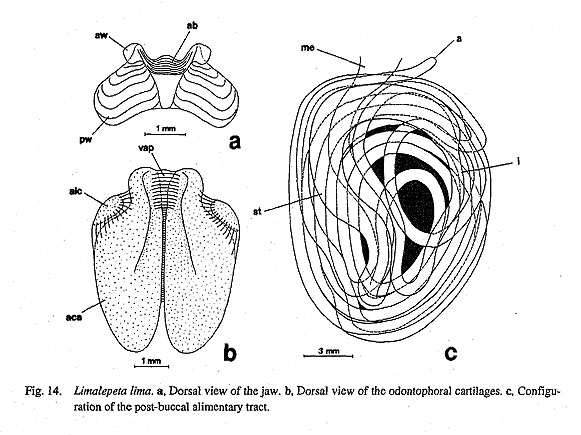
Radular formula 2-0-1-0-2. Radular row symmetrical. Central tooth paddle-shaped, nearly square, attached to radular membrane in smaller area; cusp divided into three portions of equal width. Marginal teeth broadly spatulate, gently curved. Attachment area of teeth on radular membrane small. Basal plates absent.
Subradular membrane firmly fixed to cartilages by retractors of subradular membrane (rsr), which are not divided into dorsal and ventral parts, but clearly shows paired condition (Fig. 13 b). Protraction of membrane caused by two pairs of protractor muscles. Median protractors (mpr) thin, extending toward posterior side of buccal mass. Configuration of right and left muscles completely symmetrical. Lateral protractors (lpr) thick, inserted on anterior ventral surface of subradular membrane, running above inner ventral protractors. Radular sac controlled only by retractors of radular sac (rrs). Median retractors of radular sac (mrs) and ventral tensor of radular sac (trs) (corresponding to those of Patella and Cellana) absent.
Odontophore containing two pairs of cartilages (Fig. 14 b). Anterior cartilages (aca) extending over whole length of odontophore, expanding laterally, connected by two layered ventral approximator (vap); ventral layer widely connecting to anterior cartilages, while dorsal layer restricted to anterior region between grooved parts of cartilages. Anterolateral cartilages (alc) small. Labial cartilages (as in Cellana) absent.
Sublingual pouch (slp) deep, lacking pouch-like projections and glandular outgrowths on sides (Fig. 13 b). Licker weakly sculptured by divaricate furrows. Radular diverticulum widely spread over odontophore, covered by sheet-like retractors of radular diverticulum (rrd, Fig. 13 a). Salivary glands arising directly from lateral walls of buccal cavity, extending obliquely toward outer posterior corners of buccal mass.
Anterior esophagus depressed over odontophore, with dorsal food channel and lateral pouches. Midesophagus (me) septate internally, not strongly inflated, with rather gradual counterclockwise rotation in this part. Posterior esophagus (pe) marked by internal furrows; its initial part not distinctly constricted.
Stomach (st) basically C-shaped, but its anterior part distorted posteriorly (Fig. 14 c). Openings of digestive glands restricted to small area of initial part. Gastric caecum, sorting area, and tooth of gastric shield absent. Intestinal groove between typhlosoles very shallow. Intestine (i) extremely long, complicated in configuration. Rectum passing pericardium along posterior wall, terminating at right corner of pallial cavity.
Circulatory System: Heart consisting of auricle, ventricle, and muscular bulbous aorta (Fig. 12 a). Auricle (au) receiving blood from efferent pallial vessel on right side and also directly from roof of pallial cavity. Ventricle (v) elongated along posterior wall of pericardium, leading ventrally into muscular bulbous aorta (ba) which has similar form to that of ventricle.
Excretory System: Paired kidneys of extremely unequal size on right side of pericardium (Fig. 12 a).
Left kidney (lk) in area between anus and pericardium; lumen filled with minute digitiform projections. Left kidney opening (lko) projected as small papilla. Course of renopericardial ducts not determined. Right kidney (rk) encircling alimentary tract along shell muscle, also widely extending into base of visceral mass; lumen spacious with membranous partitions. Right kidney opening (rko) extended as long tapering tube (Fig. 12 b).
Reproductive System: Gonad lying on ventral side of digestive tracts and glands. Gonoduct arising from left side of gonad, communicating with right kidney anteriorly, passing below rectum; relationship with right renopericardial duct not determined. Right excretory pore prolonged as urogenital opening.
Nervous System: Circumesophageal nerve ring hypoathroid. Pleural and pedal ganglia very closely connected. Right and left pedal ganglia widely spaced. Labial ganglia united by labial commissure. Buccal ganglia arising from posterior sides of labial ganglia. Visceral loop originating from left and right pleural ganglia. Supraesophageal, visceral, and subesophageal ganglia fused into tight loop behind buccal mass. Osphradioctenidial ganglia and zeugoneury absent. Pedal cords widely spaced. Statocysts lying on outer sides of pedal ganglia below pleural ganglia.
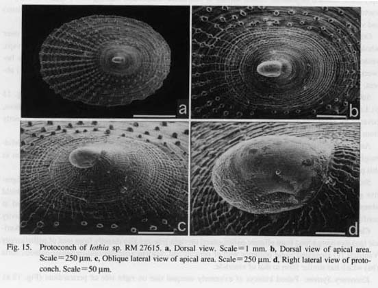
lothia sp.
(Figs. 15 a-d)
Protoconch: Protoconch completely symmetrical, longitudinally elongated. Lateroventral wall extending outward to form lateral pouches and corresponding constrictions on either side. Densely reticulated sculpture and several longitudinal streaks visible on side, although detailed structure not observed in specimens examined due to erosion. Protoconch symmetrically positioned relative to sagittal axis of teleoconch throughout ontogeny.
Family Acmaeidae Forbes, 1850 Pectinodonta orientalis Schepman, 1908
(Figs. 16 a-d, 17a-e)
External Anatomy: Mantle margin (mm) apparently lacking definite microtentacles in preserved specimens; living specimens not observed. Shell muscle (sm) horseshoe-shaped, constricted into about eighteen bundles. Head with short snout and non-papillate cephalic tentacles (ct) without eyestalks. Outer lip of mouth surrounded by oral lappets. Presence or absence of eyes not ascertained. Epipodial region smooth, lacking sensory streaks on shell muscles. Epipodial tentacles and sense organs, and operculum also absent.
Pallial Complex: Pallial cavity shallow, restricted to nuchal region, containing left ctenidium, paired kidney openings, and anus (Fig. 16 a). Osphradium and hypobranchial gland absent. Feather-like bipectinate ctenidium (c) arising from left side in front of pericardium. Ctenidial membrane not developed on both afferent and efferent sides. Ctenidial lamellae lacking sense organs and skeletal rods.
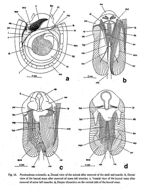
Digestive System: Inner lips of mouth (il) pulled outside by transverse labial muscles (tlm). Oral tube not surrounded by buccal sphincter and constrictor. Muscles of odontophore include the following (Figs. 16 b-d): (1) Dorsal protractors (dp) inserting on posterior end of posterior cartilages. (2) Lateral protractors (lp) occurring anteriorly on posterior cartilages, extending to lateral walls of snout and posterior wings of jaw; insertion slightly overlapping ventral protractors. (3) Outer ventral protractors (ovp) as most prominent muscle on ventral side, covering entire ventral side. Inner ventral protractors (ivp) inserted on posterior cartilages, connecting to posterior end of sublingual pouch (slp), ventral side of oral tube, and anterior wings of jaw (aw). (4) Anterior levators (al) on dorsal side between strands of dorsal protractors inserted on anterolateral cartilages (alc). Depressors and postdorsal and dorsal buccal tensors absent.
Jaw situated posterolaterally over oral tube, composed of anterior and posterior wings (aw, pw) connected by smooth anterior band (ab) (Fig. 17 c); posterior wings attached to anterolateral projections of anterior cartilages.

Radular sac not very long, forming two loops behind buccal mass, posterior end not bifurcated. Radula formula 0-1-0-1-0. Radular row symmetrical. Lateral teeth widely lobate, prominently serrated with ten denticles. Cusp divided by faint line into two portions, i.e. innermost and remaining denticles. First denticle most prominent, with remaining ones becoming shorter distally. Basal plates very solid with oblique tooth attachment scars; anterior margin concave; posterior margin convex; anterior outer corner projecting anteriorly, articulating with posterior margin of adjacent plates.
Muscles of subradular membrane consisting of the following (Fig. 16 d): (1) Median protractors (mpr) very thin, extending to ventral body cavity posterior to buccal mass. Both right and left strands running straight, exhibiting symmetrical configuration (unlike Patella and Cellana). (2) Lateral protractors (lpr) attached to subradular membrane on inner tip along with median muscle, obliquely extending posteriorly to origin of posterior cartilages. Muscle occurring just beneath inner ventral protractors (ivp). (3) Subradular membrane widely attached dorsally by retractors (rsr) that constitute main part of buccal mass. Right and left halves clearly divided into paired condition, not fused ventrally (unlike Patella and Cellana). Ventral side of radular sac connected to posterior cartilages by retractors of radular sac (rrs). Radular sac manipulated only by retractors of radular sac. Median retractor and ventral tensor of radular sac absent.
Odontophoral cartilages consisting of larger anterior and smaller anterolateral cartilages (aca, alc) (Figs. 17 a, b). Anterior pair united by two-layered ventral approximator (vap).
Sublingual pouch (slp) provided with triangular sac on sides (lsp) (Fig. 16d); wall very thin, probably not glandular. Licker not sharply lamellate but sculptured by furrows. Radular diverticulum (rdv) widely spread over odontophore. Salivary glands obliquely extending from lateral part of buccal cavity toward outer posterior comers of buccal mass. Salivary ducts absent.
Anterior esophagus dorsoventrally depressed, divided into median dorsal food channel and lateral
esophageal pouches on outer sides by dorsal and ventral folds. Inner wall of midesophagus densely septated. Dorsal and ventral folds twisted counterclockwise internally in this section. Posterior esophagus becoming narrower, directed to right side.
Stomach with complexly folded configuration (Fig. 17 d). Proximal and distal sections curving once or twice below C-shaped dorsal median part. Openings of digestive glands very narrow, restricted to initial part of stomach. Gastric caecum, corrugated sorting area, and tooth of gastric shield absent. Intestinal groove not distinct. Intestine coiled three times, terminating at anterior right corner of visceral mass after running along posterior margin of pericardium and left kidney (Fig. 17 e).
Circulatory System: Pericardium (pc) posterior to base of ctenidium, longitudinally elongated into triangular form (Fig. 16 a). Heart consisting of single auricle and ventricle. Auricle (au) connected to efferent pallial and efferent ctenidial vessels. Ventricle (v) larger and more extensively muscular than auricle, opening ventrally into muscular bulbous aorta through long slit. Afferent ctenidial vessel connected to blood space of left kidney. Efferent ctenidial vessel (ecv) emptying directly into auricles together with efferent pallial vessel (epv) from circumpallial vessel.
Excretory System: Two extremely unequal kidneys on right of pericardium. Left kidney (lk) much smaller than right. Left kidney opening (Iko) non-papillate, consisting of small transverse slit. Renopericar-dial ducts not observed. Right kidney (rk) encircling digestive system dorsally and ventrally. Right kidney opening (rko) projecting forwards as papilla.
Reproductive System: Gonad lying ventral to digestive tract and glands. Gonoduct arising from left side, connecting to right kidney.
Nervous System; Three major ganglia of circumesophageal nerve ring hypoathroid. Pleural-pedal complex concentrated. Pedal ganglia almost fused in midline. Labial ganglia present; labial commissure below sublingual pouch. Pedal cords very thick. Statocysts on outer sides of pedal ganglia.
Family Lottiidae Gray, 1840
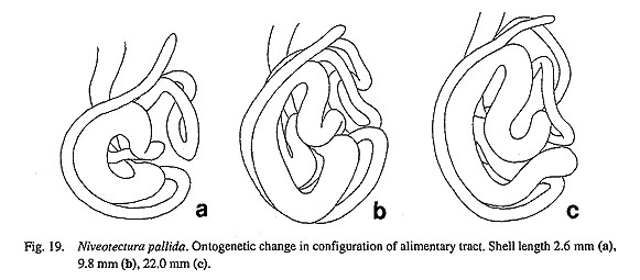
Niveotecturapallida (Gould, 1859)
(Figs. 18a-c, 19a-c)
External Anatomy: Pallial tentacles on edge of mantle margin present in living condition, but so minute and sparse that they can be hardly observed in preserved specimens. Shell muscle horseshoe-shaped, very thick, divided into approximately fourteen bundles by blood penetration. Head with pair of thick nonpapillate cephalic tentacles (ct) without eyestalks. Mouth encircled by oral lappets, smooth, without micro-tentacles. Eyes (e) deeply pitted, black-pigmented within bases of cephalic tentacles. Epipodial part lacking tentacles and sense organs. Operculum absent. Pallial streaks absent on shell muscle.
Pallial Complex: Shallow pallial cavity contains left ctenidium, paired kidney openings, and anus (Fig. 18 a). Osphradium and hypobranchial gland absent. Ctenidium (c) triangular, not very long. Ctenidial membrane not developed. Lamellae lacking sense organs and skeletal rods.

Digestive System: Inner lips connected to transverse labial muscles on inner and ventral sides. Odonto-phore narrower in anterior half, expanded laterally in posterior region, fixed by three pairs of protractors and one pair of levators (Fig. 18 b). (1) Dorsal protractors (dp) originating from dorsal wall of snout covering posterior wings of jaw, running along radular sac and esophagus, inserting on posterior cartilages. (2) Lateral protractors (lp) arising from outer ventral sides of, and attached to, anterolateral sides of buccal mass. (3) Ventral protractors composed of thick muscles covering major part of most ventral area of buccal mass, extending to posteroventral end of posterior cartilages. (4) On dorsal side, anterior levators (al) suspending buccal mass dorsally, originating from ventrolateral part of anterolateral cartilages, partly overlapping with inner section of lateral protractors and dorsal protractors. Depressors, and dorsal and postdorsal buccal tensors absent.
Jaw with well-rounded anterior and posterior wings (aw, pw) and smooth anterior band (ab). Anterior wings of jaw connected to inner lips. Posterior wings fixed to odontophore by muscular attachment from anterolateral cartilages.
Radular sac short, forming two small loops, with posterior end simply rounded. Radula formula 0-3-0-3-0. Radular row symmetrical. Lateral teeth arranged in inverted V-shape. Blunt cusp of each tooth strongly bent straight backward. Innermost laterals on anterior inner sides. Middle laterals rectangular in frontal view, wider than innermost teeth distally. Middle and outermost teeth continuously situated postero-laterally. Basal plates much wider than long.
Anterior area of subradular membrane connected to paired thin median and thicker lateral protractors (mpr, lpr). Subradular membrane attaching to thick bundles of retractors. Median protractors with symmetrical configuration; retractors of subradular membrane not fused ventrally. Ventral side of radular sac directly ded to inter posterior portion of posterior cartilages by its retractors. Median retractor of radular sac and ventral tensor of radular sac absent on ventral side ofradula sac.
Odontophore containing two pairs of cartilages. Anterior cartilages (aca) inflated, broadened posteriorly,
connected by two-layered ventral approximator (vap). Anterolateral cartilages (ale) small and rounded (Fig. 18c).
Sublingual pouch deep and smooth. Outgrowths of sublingual gland and sac-like projections absent on sides of sublingual pouch. Radular diverticulum very widely spread over odontophore. Licker sculptured by several V-shape clefts. Salivary glands obliquely elongated, directly opening into buccal pouch without duct.
Anterior esophagus partitioned into dorsal food channel and lateral pouches by thick dorsal and ventral folds. Ventral folds subdivided by two longitudinal grooves at proximal portion of mid-esophagus. Both dorsal and ventral folds clearly twisted counterclockwise. Wall of mid-esophagus bearing numerous transverse lateral folds. Posterior esophagus narrowing in posterior region, distinguished by bands of longitudinal muscles.
Configuration of stomach and intestine becoming increasingly complicated with growth (Figs. 19 a-c), Stomach initially C-shaped, changing with growth to coil in posterior terminal part, finally exhibiting at least five loops. Digestive glands opening into stomach through small paired pores at beginning of stomach. Gastric caecum and tooth of gastric shield absent. Sorting area not clearly formed. Typhlosoles and intesti- nal groove ill-defined. Intestine forming numerous small irregular loops which fill large space of visceral region (Fig. 19 c). Rectum not penetrating pericardium or ventricle.
Circulatory System: Heart consisting of unpaired auricle, ventricle, and bulbous aorta (Fig. 18 a). Auricle (au) triangular, narrowed posteriorly, connected to anterior end of pericardium and ventricle posteriorly, receiving efferent pallial vessels, capillary vessels of nuchal cavity roof, and efferent ctenidial vessel. Ventricle (v) well-expanded, attached to pericardial wall on right and posterior comers, suspended by connective tissue from pericardial wall and roof. Bulbous aorta (ba) swelling beneath ventricle, dividing into anterior and posterior aortae.
Excretory System: Two extremely unequal kidneys on each side of rectum (Fig. 18 a). Left kidney (lk) much smaller than right, connected to pericardium by renopericardial canal. Right kidney (rk) surrounding stomach, broadly spreading over base of visceral region. Right renopericardial duct not found. Right opening of kidney papillate; while left opening (lko) represented by simple, transverse slit.
Reproductive System: Gonad in ventral part of visceral region, extending thin gonoduct to right kidney. Red ovary clearly distinguishable in color from creamy testis.
Nervous System: Circumesophageal nerve ring hypoathroid. Cerebral ganglia lying laterally near buccal mass at bases of cephalic tentacles, giving connectives laterally to labial ganglia and posteriorly to pleural and pedal ganglia. Cerebral commissure running in front of jaw. Labial ganglia embedded in ventral part of mouth, innervating lips of mouth. Labial commissure connecting labial and cerebral ganglia.
Right pleural ganglion connected to supraesophageal ganglion at left; left pleural ganglion associated with subesophageal ganglion at right. Visceral ganglion lying over anterior aorta, fused with supraesophageal and subesophageal ganglia to form narrow visceral loop. Visceral ganglion innervating pericardium, digestive glands, and tract. Subesophageal ganglion supplying branches to kidney and right side of shell muscles. Supraesophageal ganglion associated with ctenidium and left side of shell muscle.
Pleural ganglia attached to ventrolateral body wall, linked with pedal ganglia by thick connective tissue, laterally giving rise to anterior and posterior pallial nerves which innervate mantle margin through shell muscle. Pedal ganglia very closely situated, extending thick pedal cords. Statocysts at ventral sides of pleural ganglia.
|