CHAPTER 7
Plant Remains from the 1984 Excavations at Douara Cave
Akiko Matsutani
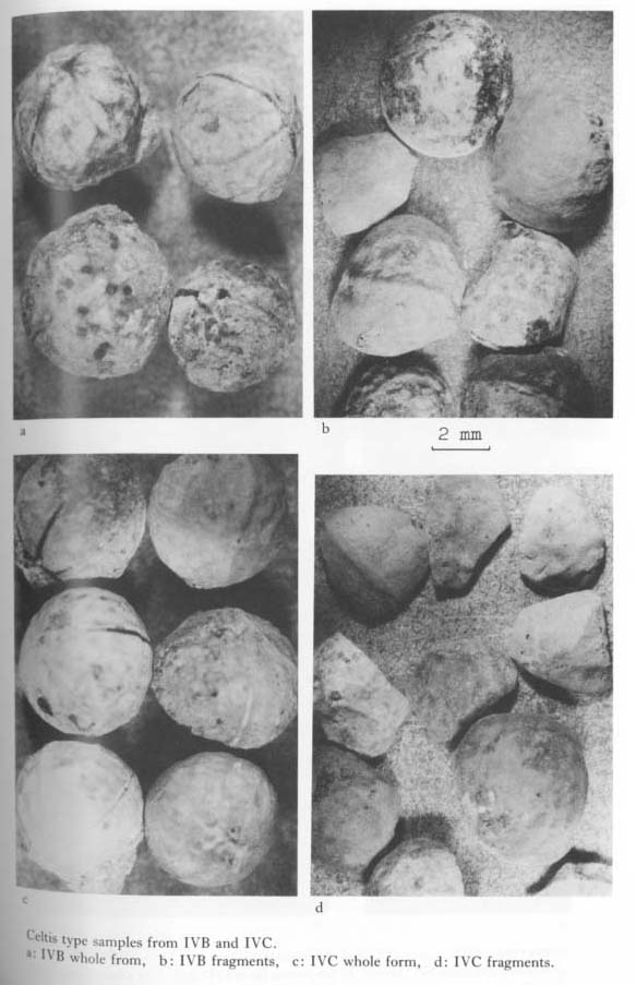
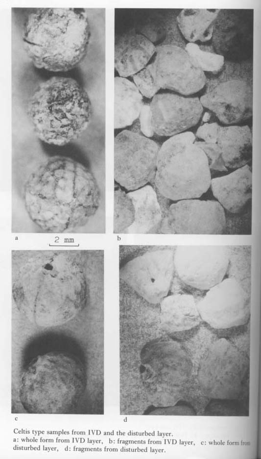
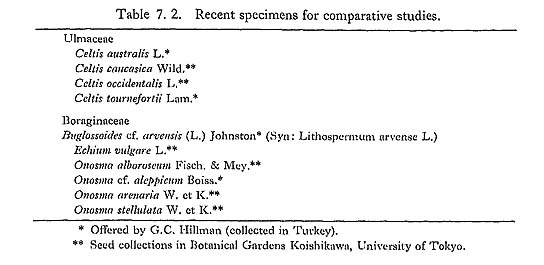
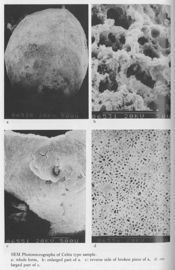
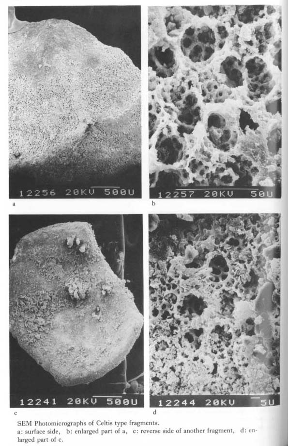
7. 1 INTRODUCTIONI have studied peculiar cell structures associated with the Levantine Mousterian deposits of the Douara Cave. Through a detailed description of the cell structure classifiable as several types (Matsutani, 1973), I have reported that it shows striking similarities to structures found in some fruits of Boraginaceae (Matsutani, 1979). In the 1984 excavations at Douara Cave, many plant remains including a number of complete specimens were found in the stratigraphic units IVB to IVD. The objective of this chapter is to identify the species of newly obtained samples in order to provide the basic data discussing environmental conditions around the Douara site. 7. 2 MATERIALS AND METHODSTwo kinds of samples were analyzed: the first involved almost complete specimens arbitrarily collected from the deposits during the excavation work, and the other consists mostly of fragmentary pieces recovered by dry-sieving method of 1 mm and 4 mm mesh in the laboratory. These samples were studied by two research methods: first, they were observed under a binocular microscope to study external morphology, and secondarily, examined for the more minute structures using scanning electron microscope (HITACHI S-700 SEM). 7. 3 RESULTS OF THE BINOCULAR MICROSCOPYThe samples examined were discriminated into three types of remains under the stereoscopic microscope. The first type is characterized by having round shape with two ridges meeting at right angles. The size is about 5-7 mm in diameter with thick walls. The surface is shallowly sculptured with net-like patterns (Plates 7. 1: a, c, 7. 2: a, c). They were brown, grey and white in color. The shape and size match wholly with fruit stones (endocarps) of some Celtis spp. Fragmentary pieces characterized by having the same features of net patterns and ridges with thick walls were also collected (Plates 7. 1: b, d, 7.2: b, d), More than seventeen samples from IVB were recognized as Celtis sp. as shown in Table 7.1 and from layer IVC more than twenty samples were also assigned to Celtis stones. However, from layer IVD, only three complete specimens together with a few fragments were collected. Besides these, ten complete stones and several fragments were collected from disturbed deposits of IVB to IVD. Three specimens from the IVD layer appeared to have more clear net patterns compared with the overlying layers samples.
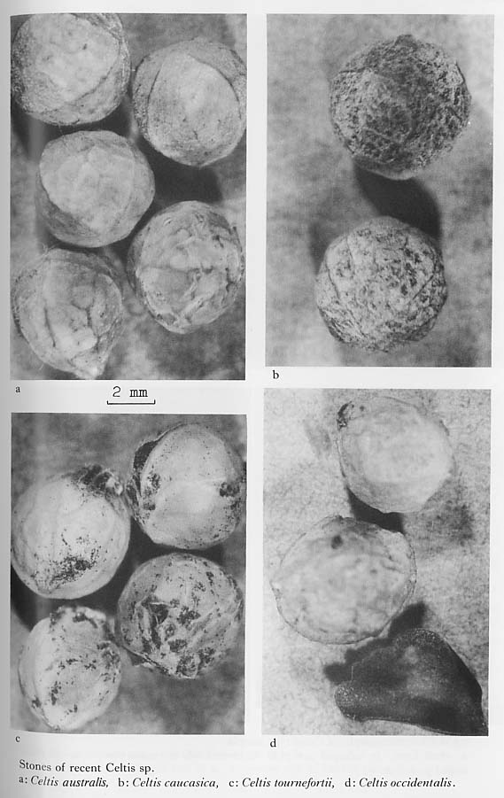
According to Mouterde (1966), Celtis anstralis and Celtis tournefortii grows widely in Syria and Lebanon. Considering the past vegetation, other species might be considered as candidates. Celtis cawasica and Celtis occidentalis were available. The identification key for these species seems to be based primarily on the nature of reticulation on the stone surface. Celtis australis has clear reticulation and Celtis tournefortii has relatively smooth surface with rough reticulation, but the other two species have an intermediate degree of reticulation (Plate 7. 3). Thus, it seems safe to divide these four species into two groups: the australis type with a reticulated structure, and the tournefortii type with a smooth surface. Through examination of the complete samples collected, it can be stated that the specimens from IVD conforms closely the Celtis australis type and that the specimen from IVB, IVC and disturbed layers are of the Celtis tournefortii type.
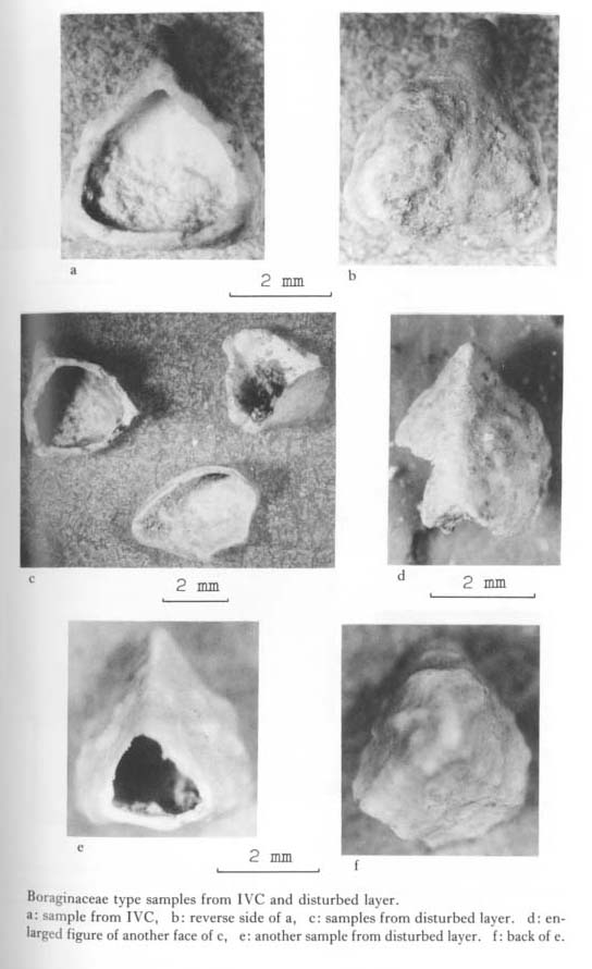
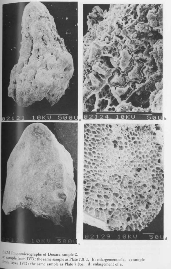
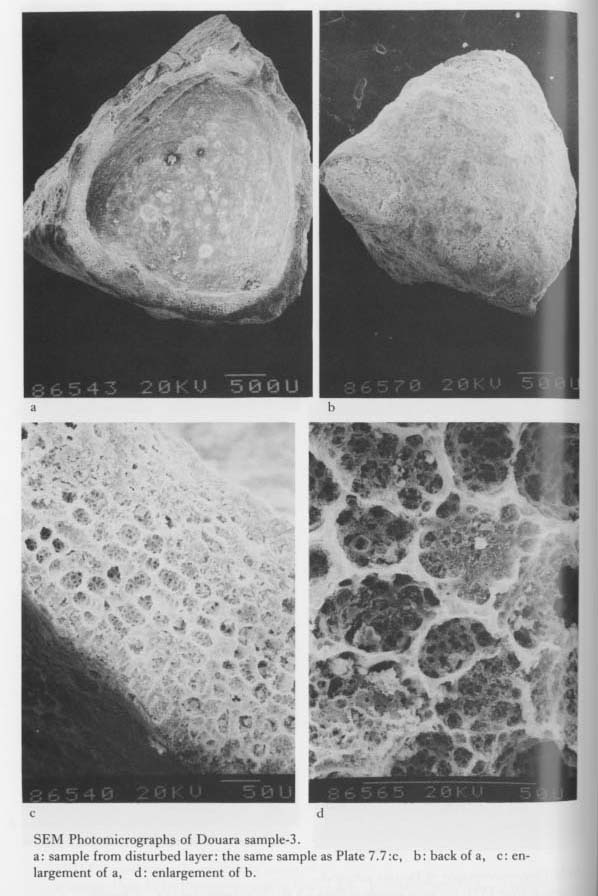
Fragmentary samples are divided into two groups in color: one white and the other grey. The former have ridges with less reticulation and with thicker section, while the latter have clear reticulation with thinner section. The former may be referred to Celtis tournefortii and the latter to Celtis australis. If these identifications are correct, the two different types are mixed throughout the deposits. The second type of mineralized remains of plants were recovered from IVC, IVD, and disturbed layers (Table 7. 1). Their occurrences were much fewer than Celtis type specimens. These were white colored and fragile with thin walls. The shape is trigonous or pyramidal (Plates 7. 7, 7. 8). The most common type is about 5 mm in height and about 4 mm in diameter. The surface is almost smooth with slight tuberculation. These features are closely related to nutlets of some Boraginaceae.
In the desiccated zone like western Asia, hundreds of species of Boraginaceae now grow (Davies, 1978). The most frequent species identified to date are three genus of Bvglossoides, Echium, and Onosma (Plate 7. 9). Comparing the Douara samples with these species, Echium is much smaller than the Douara samples, One of the most small-sized samples from IVD (Plate 7. 8: c) resembled the Buglossoides arvensis. Onosma species are frequently grown in the western Asia and their nutlets vary from small to large in size. Compared with available recent samples, Onosma aleppica (Plate 7. 9; d) showed striking similarities to the Douara ones, but it was almost twice as large. Onosma alborosea (Plate 7. 9: e) is almost the same size as the Douara specimen but its shoulders of lateral sides are rather rounded, not angular. On the contrary, Buglossoides arvensis (Plate 7. 9: a, b) has also very similar shape to the Douara collection, but it is only half the required size.
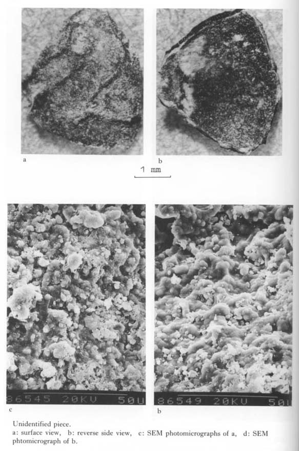
The third type is black in color with thin wall, but hard crusted. They have large netlike ridges (Plate 7. 18). It cannot be identified as any definite species.
7. 4 SEM OBSERVATION OF PLANT REMAINS7. 4. 1 SEM observation of Celtis type samplesTwo samples from IVC (Plates 7. 4, 7. 5) were observed by SEM after the following procedures.
Being washed by acetone, the samples were mounted on the stub with double scotch tape, and were coated by Pt. ion, and then observed with 20 kv. currents. One sample (Plate 7. 4) retained complete form with slight reticulation and the other (Plate 7. 5) remained almost half part with clear reticulation. The reticulation was not so clearly observed from the surface of the samples and only two ridges were seen to cross. In places circular cells of about 25 microns diameter were observed below the surface, Each cell was slightly sunken and was filled with several circular holes (Plates 7. 4: b, 7. 5: b). All over the reverse surface of the complete sample were covered with fine pitted cells and in places circular breaks existed (Plates 7, 4: c, 7. 5: c). The size of these features matched wholly with those of the circular cells of the outer surface (Plate 7. 4: d), The reverse side covers of the half-sized sample were broken and disclosed circular cells which also had sunken holes (Plate 7. 5: d). The surface and the reverse side of fragmentary samples also disclosed almost the same structures mentioned above (Plate 7. 6), and also the same structures were observed on the surface of complete specimens.
7. 4. 2 SEM observation of Boraginaceae type samplesSome samples classifiable as Boraginaceae, which held almost whole in form, were submitted for SEM observation (Plates 7. 10-12). There were three types according to the size. The first, smallest sample from IVD had an overall shape and size similar to Buglossoides arvensis, Echium vulgare, and Onosma stellulata. The sample had zigzag cells in form along the central axis (Plate 7. 10: b). Another sample morphologically similar but not the same in shape, has some pitted cells below the surface (Plate 7. 11: a, b).
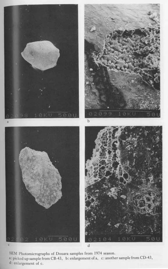
The second sample from IVD was fragmentary and apparently trigonous in shape. The specimen revealed polygonous cells with net-like pitting in the section and in hypodermis. This type of sample is more frequently encountered in IVC and the disturbed layers. The sieved sample from IVD might also be of this type (Plate 7. 11: c, d). The third sample from IVC layer, which is more fragmentary, is supposed to be larger than the former two. This specimen revealed another different cell pattern: more deep degree of zigzag shape of cells interlocking with neighboring cells as it looks like pieces of zigsaw puzzles (Plate 7. 10: c, d). The same type of structures was detected in some fragmental samples from the 1974 excavations (Plate 7. 13: b).
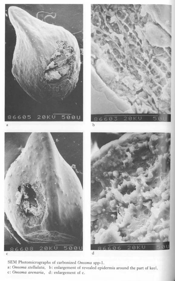
7. 4. 3 Comparison with nutlets of recent BoraginaceaeThree genus (Echium, Buglossoides, and Onosma) were selected to be compared with the excavated samples based upon the gross morphology of nutlets. The comparative specimens were carbonized, and observed by SEM after coating with Pt. ion. Echium vulgare (Plate 7. 14) is similar in shape to the second type, but it is only half the required size. The microstructures do not resemble. Buglossoides arvensis of both tuberculate and smooth types were then compared. The latter type resembled to the second type both from outer shape and from microstructure (Plate 7. 15). But it was only half the size of the Douara specimens. As noted before, Onosma spp. are variable in size. Although Onosma stellulata matches with the small complete sample from IVD, and as regards to the hypodermal cells they did not resemble (Plate 7. 16).
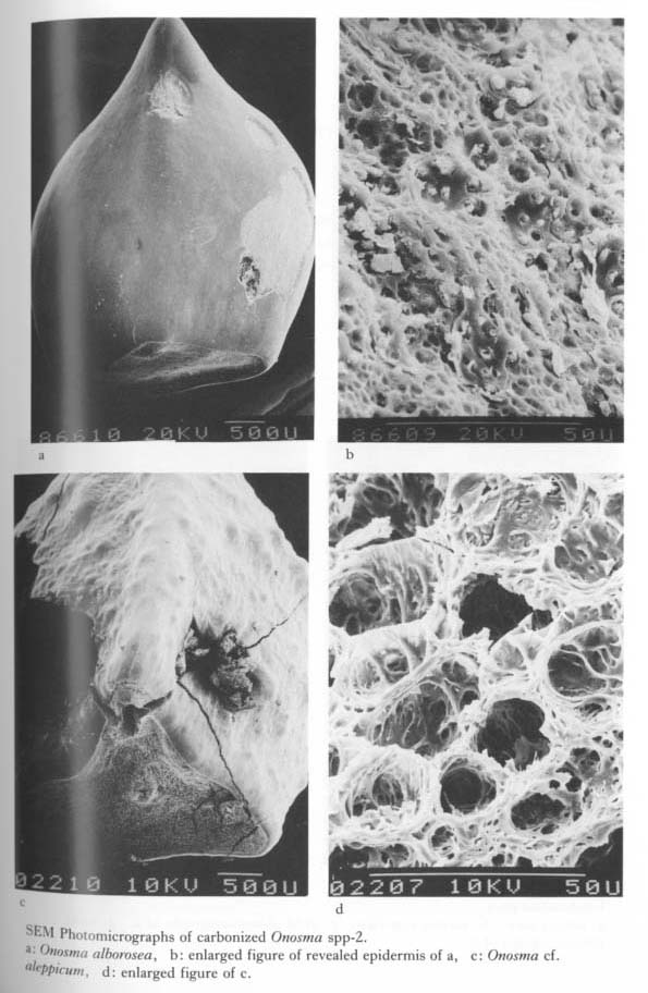
Onosma arenaria (Plate 7. 16: c) is larger than the smallest specimen and smaller than the second type. As regards to the hypodermal cells, the frims of the circular cells are notched and inside of them were existed small pitting (Plate 7. 16: d). Onosma alborosea (Plate 7. 17: a) is almost the same size as the second type and the carbonized sample easily revealed the underlying cells (Plate 7. 17: b). Onosma cf. aleppicum (Plate 7. 17: c, d) was too large as noted, but had similar structure to the sample. In conclusion, it cannot be denied that a part of the plant remains from Douara is morphologically closely related to some Onosma sp. or spp.
7. 4. 4 SEM observation of the other typeThe other samples characterized by showing black-color specimen with hard and thin crust were submitted for SEM observation (Plate 7. 18). However, the species with structures comparable with the Douara specimens were not found. 7. 5 DISCUSSION AND CONCLUSIONFrom the total results described above, I found that the Douara material is composed of at least two groups of plants: one is Celtis cf. australis and/or Celtis cf. tournefortii, and the other some species of Boraginaceae. The latter had already been suspected from amorphous silica pattern of the 1974 excavations' material. Thus, this suspection has been clearly evidenced by the present study. Celtis sp. are widely grown for edible fruits. In the field of prehistory, stones of Celtis sp. were found in ash deposits of Choukoutien Cave in China which had been inhabited by Peking Man some 500 thousands years ago or so (Brothwell and Brothwell, 1969). Stones of Celtis sinensis were also reported at prehistoric sites in Japan (Kokawa, 1980). In western Asia, Celtis sp. has been identified at a number of Neolithic sites such as Cayönü (Van Zeist, 1972), Hacilar (Helbaek, 1970), Can Hasan III (French et al., 1972) in Turkey, and Tell Abu Hureyra (Hillman, 1975) in Syria. The usage of this fruit is said to be material for wine (Renfrew, 1975). These Neolithic sites, in addition to Celtis sp., produced some borage seeds, although the usage of these plants proved to be puzzling. Lithospermum arvense and Heliotropium europaeum with Celtis australis (Hacilar), Amhusa and cf. Boraginaceae with Celtis tournefortii (Cayönü), Lithospermum arvense and Echium sp. with Celtis tournefortii (Tell Abu Hureyra), cf. Lithospermum arvense with Celtis cf. tournefortii (Can Hasan III). Seed of Boraginaceae not accompanied by Celtis stones were found from other sites as Beidha in Jordan (Helbaek, 1970), Ali Kosh in Iran (Helbaek, 1969) and Franchthi Cave in Greece (Hansen, 1978). It is to be noted that the quantity of these two groups were found far more greater than other species. This evidence may be concerned with preservation condition of the material due to the abundance of silica and calcium in the cell walls of these groups. So it is not to be deduced that these two groups of plant were highly evaluated by the Douara people. Perhaps many other species such as were listed in the other sites in western Asia might had contributed to the useful plant list though not survived nor remained at present. However, the question still remains what part of role the borage family played for the Douara people. In the previous study (Matsutani, 1979), I considered the possibility as food based on the find of Borago officinalis from stomach by Netolitzky (1912). But now I am inclined that about the usage of this family by the Douara people the roots was used for dye and the nutlets together with leaves and stems were residue or waste, so they were discarded after extraction of chemicals and used for fuels together with Celtis stones after eating edible part. Lastly I must make some remarks on the connection to the amorphous silica. In the previous study, fruits of some species of Boraginaceae were incinerated and were compared with the amorphous silica. Then Borago officinalis, Symphytum officinalis, Pentaglottis sempervirens and Echium vulgare revealed the similar patterns with heavy cell. These results did not accord with the identification from outer morphology revealed this time except Echium. One explanation to this fact may be that the latter species such as Buglossoides and, Onosma were more hard-crusted than the former, so that the amorphous silicas were composed of only the former. To attest or inspect this hypothesis, I must make clear the following three problem: A, the amorphous silica from soils and ash in the 1984 excavations must be analyzed; B, the spodograms of the latter (Onosma spp.) must be compared with amorphous silica in 1974 and in 1984; and C, leaf portion of especially traces of hairs or trichomes of above mentioned species which I did not conduct in the previous study must be done and be compared. If the part of amorphous silica might match with the epidermis of Boraginaceae, the above mentioned hypothesis might be evidenced. AcknowledgmentThe author expresses her great thanks to Dr. Gordon Hilllan, Institute of Archaeology, London University for his generous provision of modern reference samples besides appropriate advice. The author also appreciates Miss Tomoko Yamashita, The University Museum, University of Tokyo for her technical support in SEM operation. LITERATURE CITED
|