CHAPTER 4
Postcranial Skeleton of the Minatogawa Man
Hisao Baba* and Banri Endo**
*Department of Anatomy, Dokkyo University School of Medicine;
**Department of Anthropology, Faculty of Science,
The University of Tokyo
| ( 3 / 7 ) |
|


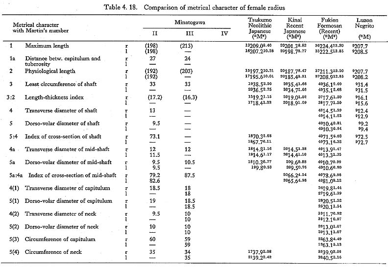  |
Caput and Collum
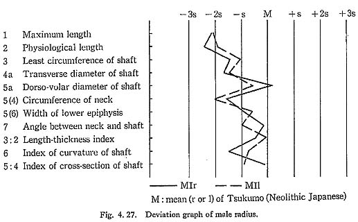
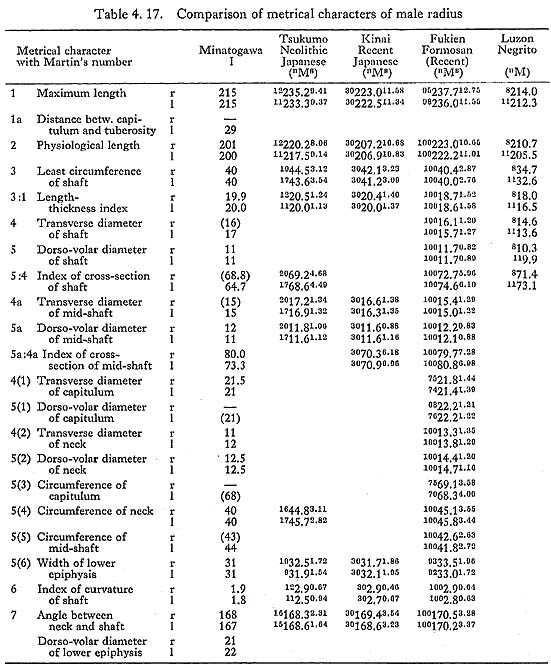
The size of the Minatogawa articular head is approximately comparable to those of the populations compared; the neck, however, seems slightly more slender. The neck-shaft angle varies from 165° to 171°, being similar to those of other populations compared. The shape of the articular head is normal.
Diaphysis
Curvature of the shaft
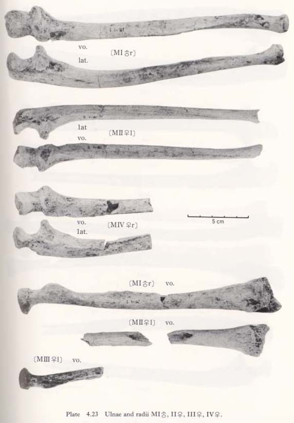
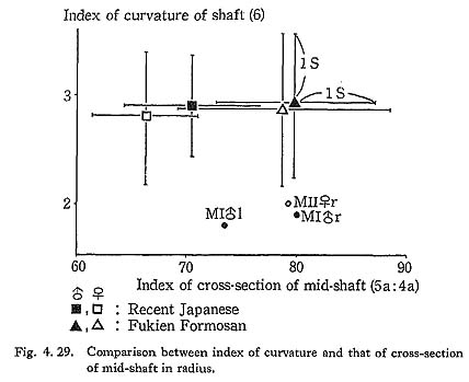
The range of the curvature index is from 1.8 to 2.2, which is rather smaller than the means of the Japanese and Fukien Formosan (Fig. 4. 29).
Marking by muscle or ligament
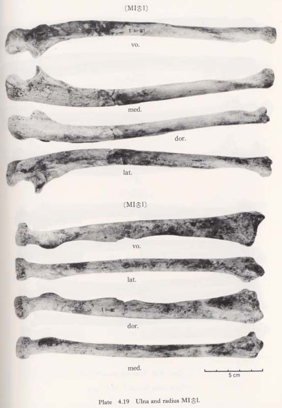
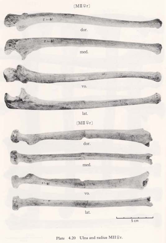
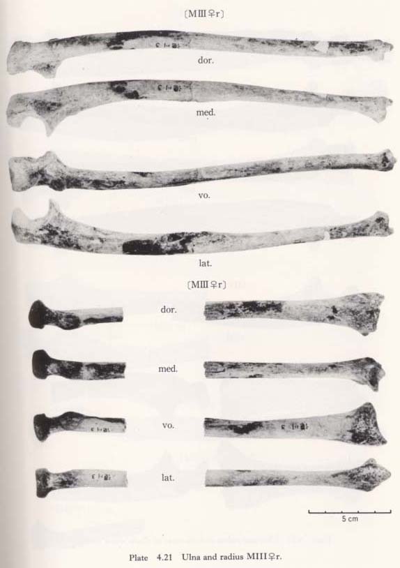
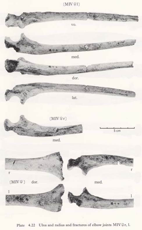
The radial tuberosity is well developed and protrudes medially in MI rl and MII
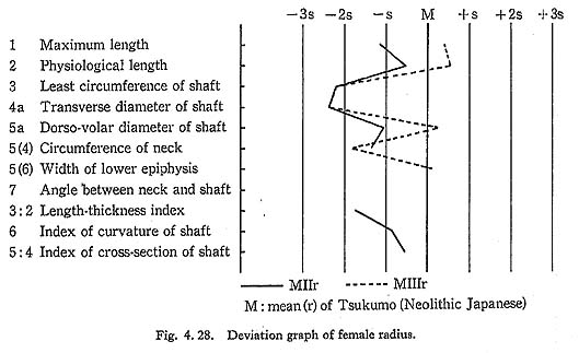
 l, whereas it is rather poor in MIIIr. The hyperostotic change is seen on the dorsal margin of this tuberosity in MIIl. The ridge for the flexor digitorum superficialis is well defined. In MIIIrl it becomes a particularly prominent crest near the radial tuberosity. On the lateral surface, the rough surface for the pronator terres is marked in the male, though poor in the females. The protruding part of the interosseus margin is high and long in MIIr, high and short in MIrl, and low and long in MIIIr. No marking is visible on the volar surface for the pronator quadratus. In MIrl and MIIIr, the depression of the distal volar surface above the carpal joint is marked in the medial half of the volar surface but is not well defined in the lateral half. It is rather poor in MIIrl.
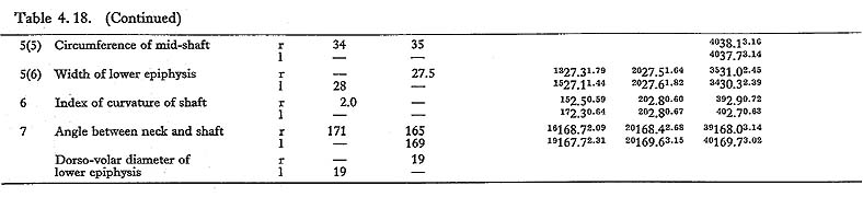
Cross-section
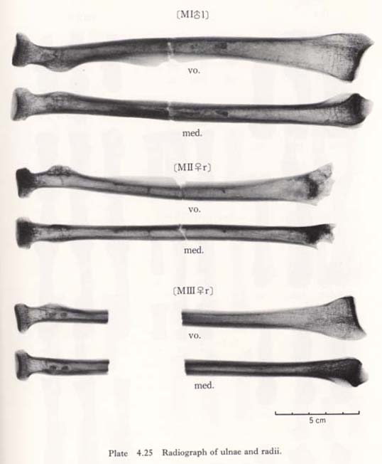
The shape of the cross-section at mid-shaft is somewhat triangular due to the development of the anterior and posterior borders in MIrl (Fig. 4. 30). It is rather flattened in MIIrl and round in MIIIr. As the cross-section index shows, the flatness of the Minatogawa radial shaft is less than that of the Japanese, and roughly equal to that of the Fukien Formosan (Fig. 4. 30).
Lower Epiphysis
The transverse diameter is slightly smaller than the mean of the Japanese. On the dorsal surface of MIrl, there is a thick ridge having a groove for the extensor pollucis longus which runs from a supero-medial to an infero-lateral direction. This ridge is also well developed but the groove is not present in MIIl and MIIIr. Laterally to this ridge, two grooves for the extensor carpi radialis longus and brevis are well distinguished, and medially to this ridge, two grooves for the abductor pollucis longus and the extensor pollucis brevis are also clearly seen in all the present specimens. No special characteristic is seen on the articular surface.
Discussion and Conclusion
Curvature of the shaft in the radius and the ulna
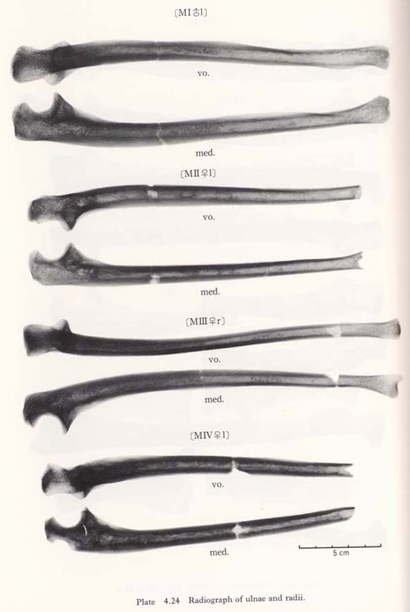
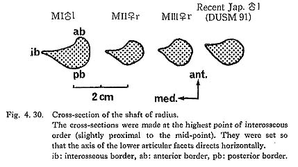
In normal cases, the ulnar shaft is straighter than the radial shaft (Figs. 4. 24, 31). On the contrary, the former is more curved than the latter in the Minatogawa forearm, As the result, the distance between these two bones or the interosseous space in Minatogawa people is roughly equal to that of usual sapiens.
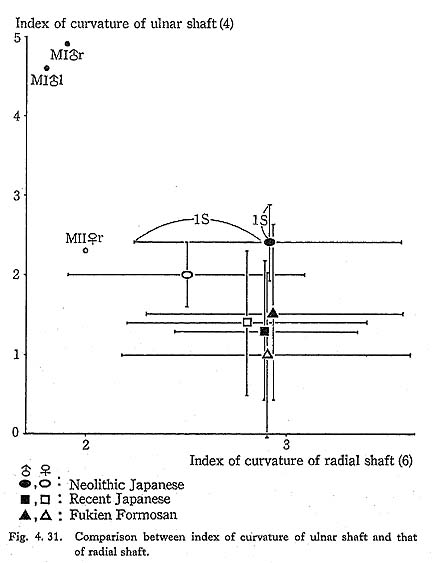
The functional meaning of this curvature is interpreted as follows. When the two bones are articulated in a semi-prone position, the ulnar convexity faces dorsally and the radial convexity faces volariy (Fig. 4. 24). In this position the axis of the combined forearm is dorsarly convex in the case of Minatogawa and volarly convex or straight in normal cases, though this difference is not marked. It is considered that the Minatogawa type of forearm is advantageous for elbow flexion, since the flexor muscles in the upper arm may produce the bending moment on the forearm which makes it convex anteriorly, while the flexor muscles may produce a reverse bending moment if the forearm convexity directs dorsarly.
Thus, as long as the flexor muscles in the forearm act simultaneously, the bending moment which acted on the forearm in the elbow flexion may have been smaller in the Minatogawa type than in the usual type.
HAND BONES
Carpus
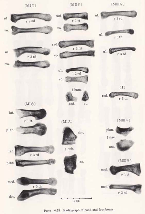
There remain two left hamates (MB1, MIIl), (Plate 4. 25). The medio-distal diameter is 17 mm in MB1, 18 mm in MIIl; the transverse diameter is 10 mm in MB1, 13 mm in MIIl. MB1 was at first classified as MI, but Minatogawa I is a stout male, so it may not belong to him, but to an unknown gracile female. MIIl is rather stout in proportion to her slender upper limb bones. The facet for the fourth metacarpal is very narrow in both specimens. The hamulus is not stout; its height is 7 mm in MB1, 8 mm in MIIl.
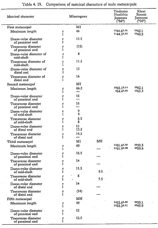
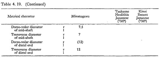
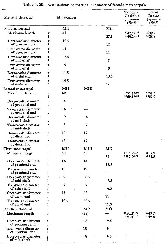
Metacarpus
There are 17 metacarpals. Of these, three are the first, four are the second, five are the third, two are the fourth and the final three are the fifth (Plates 4. 26-28).
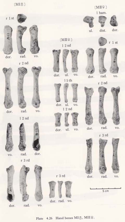
First metacarpal
MIr: male, complete. MIIr: female (?), nearly complete. MC1: female, complete, formerly classified as MI.
The maximum length is 46 mm in MIr, considerably larger than the mean of the Japanese males (Table 4. 19). It is 41 mm in MIIr, comparable to those of the other females studied (Table 4. 20). The value for MCl is smaller than that of the Japanese females. Generally, MIr seems to be stout and MIIr also stout for a female, though MCI is gracile. The area of the opponens pollucis becomes a sharp ridge in MIr. The facet for the abductor pollucis longus is well defined in all three bones. On the postero-volar margin of the distal facet in MIr and MIIr, an exostosis can be seen.
  |
  |
Second metacarpal
MIr: male, complete. MIl: male, distal two-thirds. MIIr: female (?), complete. MIIIr; female, distal half.
MIr is longer than the Japanese bones. The length of MIIr falls into an intermediate position between the means for the Japanese males and females. On both sides of the dorsal surface and along the median line of the volar surface, the crests for the interosseous muscles are well developed in all the specimens. The facet for the extensor carpi radialis is also visible in MIr and MIIr.
Third metacarpal
MIr: male, almost complete, some part of the distal end missing.
MIIr: female (?), complete. MIIIr: female, complete. MD1: female, less complete, part of the proximal end broken, formerly classified as MI, ME1: male, shaft only.
MIr and MD1 are roughly similar in length to the Japanese bones. MIIIr is very long for a female, but is proportional to her long ulna. MIIr is also of intermediate size, both in length and thickness, between the means of the males and females. MIr seems stout, having a high ridge for the ventral interosseous muscles. Its facet for the second metacarpal fits well to the corresponding facet on the second metacarpal.
Fourth metacarpal
There are two isolated bones:
MFr: female (?), distal end is broken off.
MGl: female, proximal half is present, formerly classified as MI.
The estimated length of MFr is 53 mm, which falls between the means for the Neolithic males and females. Its stoutness is roughly comparable to the third metacarpal of Mil and more robust than MGl. This bone may belong to a male. MGl surely belongs to a female, judging by its small size. Its distal end fits well to the corresponding facet of the hamatum (MB1). In addition, both are very small, even for a female, so it is considered that these two belong to the same individual.
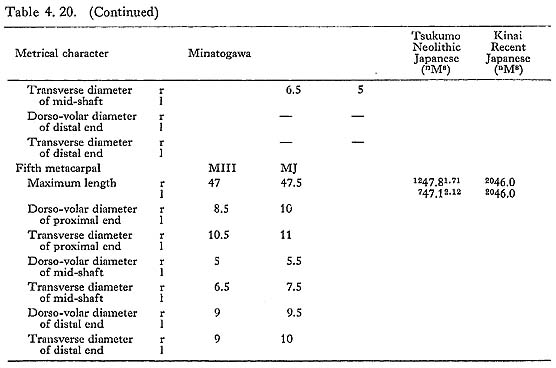
Fifth metacarpal
There are three bones.MIIIr: female, complete. MHr: male, most of the distal end missing.
MJr: female (?), complete.
The length of these three bones are not markedly different. As a male, MHr is rather short but stout. As a female, MIIIr is moderate in both length and thickness. Since MJr is intermediate between the two in size and in appearance, its sex is not clear.
Phalanges
Proximal phalange
There are six proximal phalanges. Two of them are from MII, one from MIII, the rest are isolated bones MK, ML, MM. The larger bone from MII is 38 mm in length, which is slightly less than the second and fourth bones of the Neolithic Japanese males and slightly larger than those of the females. Its proximal end is wide (14 mm). From a dorsal view, a slight inclination to the left side is seen in the distal facet, and the proximal facet is dislocated toward the left (Plate 4. 26). Considering these facts, this bone may be of the left second proximal phalange. The smaller one from MII is short (30.5 mm) and slender. The distal articular facet inclines strongly to the right side from a dorsal view. Therefore, this bone probably belongs to the left fifth proximal phalange.
MIII is long (43.5 mm), comparable to the length of the third bone from Neolithic Japanese males. However, it is relatively slender, its proximal width being only 13.5 mm. No inclination is seen on either end. Since Minatogawa III has a long ulna and metacarpals, this bone may belong to the right third proximal phalange of this female individual.
The isolated MK is rather short (33.5 mm) but very stout with a wide shaft. The inclination of the distal end is marked. It directs toward the right when dorsally seen. This bone might belong to the male left fifth proximal phalange. ML was at first classified as MI. However, because of its small size (26 mm in length, 12 mm in proximal width) it may be the female first proximal phalange. Judging from the ridge on the ventro-ulnar margin of the proximal shaft, it is from the left side. MM is rather stout and its proximal end is broken off. Its estimated length (41 mm) matches those of the third bone of the Neolithic Japanese females and the second and fourth bones of Neolithic Japanese males. A slight inclination of the distal facet is seen to the right side from a dorsal view. The shaft is straight, having no increase of transverse diameter near the proximal end, as is seen in the second bone. Hence it may belong to the male left fourth proximal phalange.
Middle phalange
There are only two bones from MI and MII. MI is 29 mm in length and 13 mm in proximal width. Its length is comparable to the third bone of the Neolithic Japanese males. It may belong to the right third middle phalange.
The length of MII is 23 mm and proximal width is 12.5 mm. Regardless of sex, this size is too small to be the third bone and also too large to be the fifth one. Accordingly, it should belong to the second or fourth middle phalange. It is rather robust and the inclination of the distal facet is to the right side from a dorsal view. Therefore it seems that this bone may belong to the left second. Its distal facet is slightly pronated from its proximal facet.
Discussion and Conclusion
Among the Minatogawa people, MI and MIII had relatively large hands; the metacarpals of MI are long and thick. Comparing the sum of the length of the first, second and third metacarpals, the value for MI is 105.5% of that for Recent Japanese males (sum of the mean values), and even a little larger than that of Neolithic Japanese males. Compared with the overall smallness of the upper limb, this large size is noteworthy.
The sum of the length of the third metacarpal and the third proximal phalange were compared for MIII. The value of MIII is 108.4% of that for Recent Japanese females, 104.9% of that for Neolithic Japanese females. It is rather comparable to those of the males. Although her forearm is rather long, this length is remarkable. On the other hand, other individuals might have had moderate or small hands proportional to the small size of their upper limbs.
Most of the hand bones of MII are very stout for a female, but the other upper limb bones indicate that MII is a small, gracile female. It seems to us that the hand bones attributed to MII do not belong to her. On the contrary, as mentioned above, MIV is a very stout female, for whom no hand bone was found. We therefore cannot neglect the probability that the hand bones of MII belong to MIV.
PELVIS
Materials
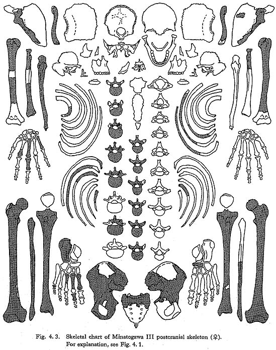
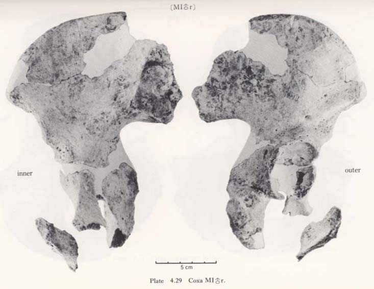
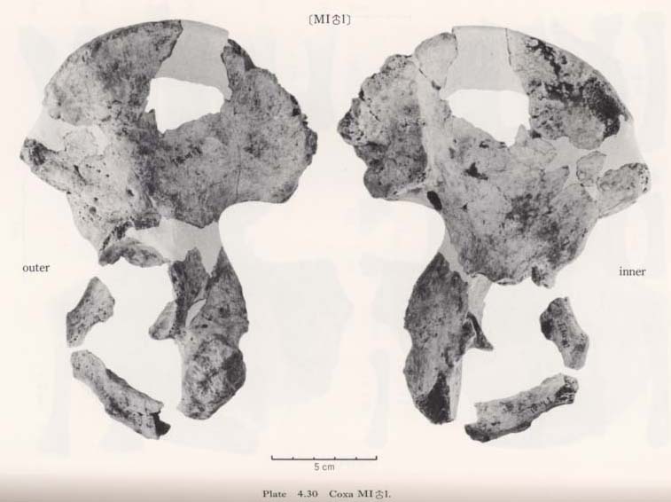
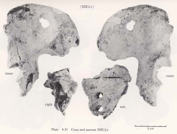
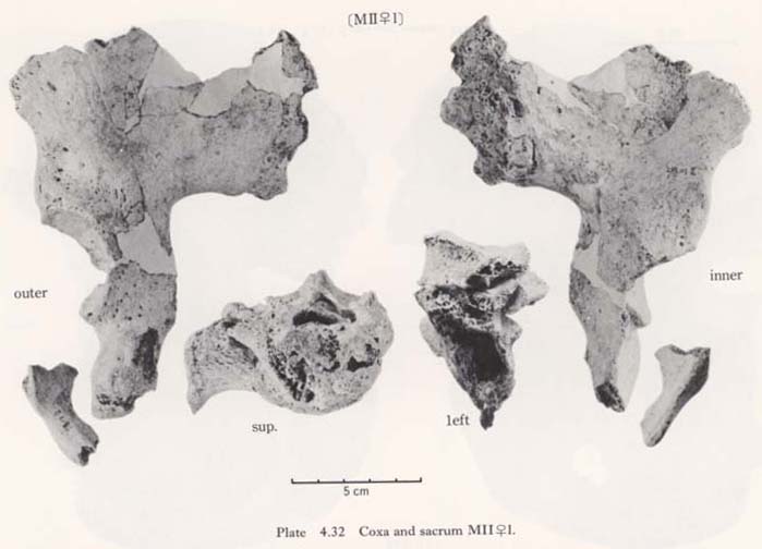
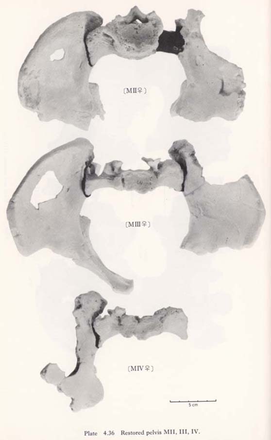
There are three pairs (MI, MII, MIII) and one right coxa (MIV) (Figs. 4. 1-4, Plates 4. 29-39).
MIr: male, most of the symphyseal region is missing and a posterior end of the ilium is broken off.
MII: male, the pubis and the ramus of the ischium are broken off.
MIIr: female, the pubis and the ramus of the ischium are broken, an anterosuperior part of the ilium is broken.
MII1: female, the upper half of the iliac wing is broken, the pubis and the ramus
of the ischium are also broken. On the auricular surface, a part of the sacrum is attached.
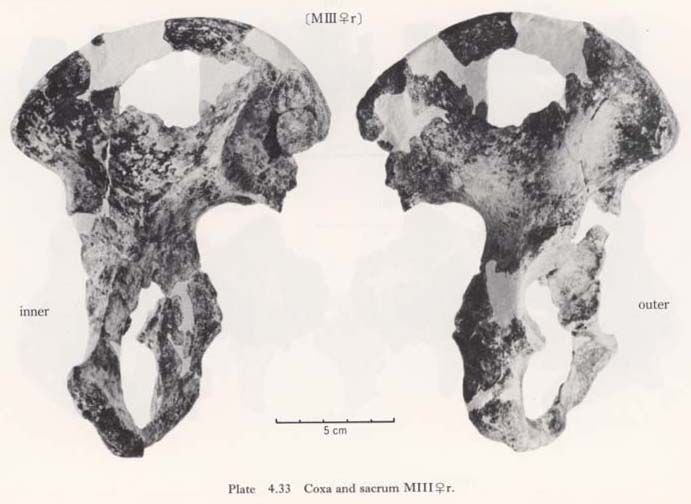
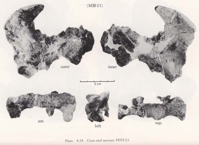
MIIIr: female, nearly complete.
MIIIl: female, only the ilium is preserved, a part of the iliac wing is broken off.
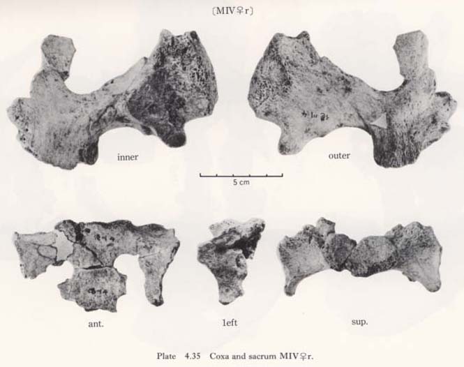
MIVr: female, a region from the acetabulum to the iliac tuberosity is present.

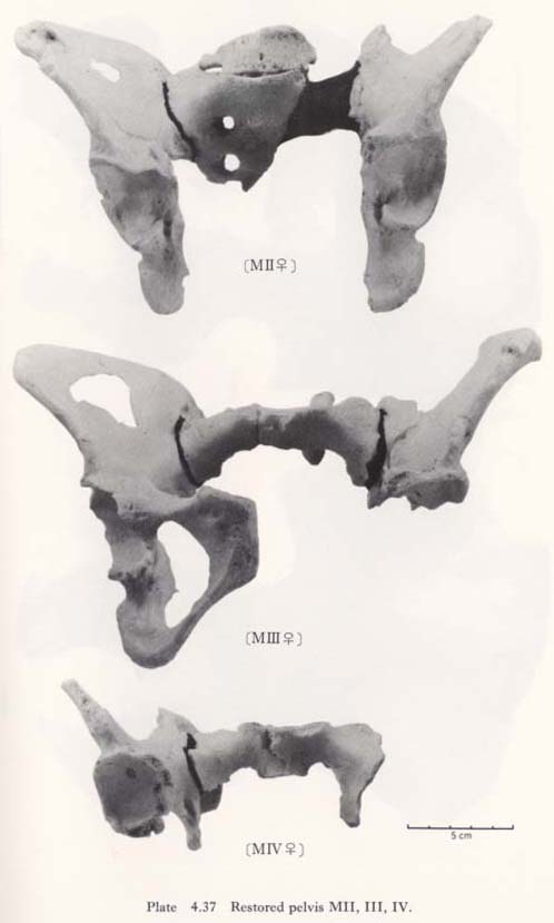
There are three sacra from MII, MIII and MIV; in addition, several flakes of sacrum from MI are found.
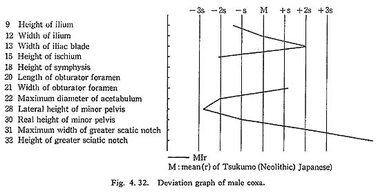
General Character of Coxa
The coxae from the Minatogawa male (I) is low and wide (Fig. 4. 32, Table 4. 21). Its maximum height is estimated to be 195 mm in MIr, considerably smaller than the mean value for Japanese males, whereas the estimated bispinal width is 156 mm, rather larger than the Japanese mean. The ratio of these two values is 80, which is quite high.
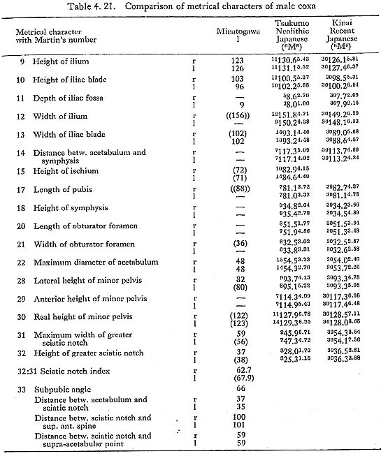
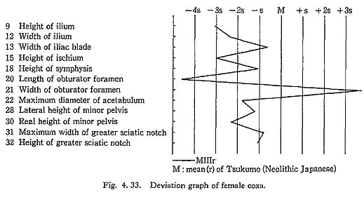
On the contrary, the restored coxae of the Minatogawa females (II, III) are low and narrow (Fig. 4. 33, Table 4. 22). The maximum height is estimated to be 176 mm in MIIr and 183 mm in MIIIr, while the Neolithic mean is 195.1 mm and the Recent Japanese females is 190.0 mm. The bispinal width is estimated as 137 mm in MIIr and 138 mm in MIIIr, both of which are roughly 10% smaller than the mean for the Japanese females. The ratio between these two items is 78 in MIIr and 75 in MIIIr, roughly equivalent to the means for the Japanese. The ratio of maximum height to femur length is 49 in MIr and MIIr and 48 in MIIIr. This Minatogawa coxa figure is slightly lower than the means for the Neolithic and Recent Japanese.
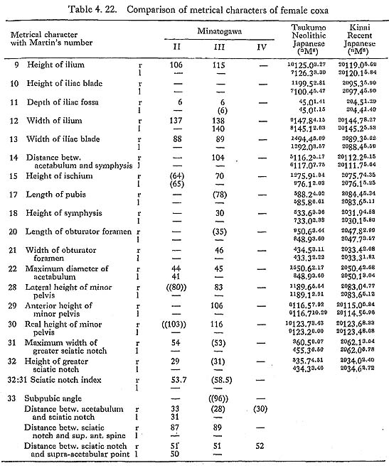
Ilium
There are four nearly complete ilia (MIrl, MIII, MIIIr) and three incomplete ilia (MIIr, MIIIl, MIVr).
General view
The values of the height of the ilium are also somewhat lower than that of the Neolithic Japanese. Proportionally MI is rather long sideways compared to its large width. MII is also long sideways compared to its small height, and MIII is moderate in its relatively small height and width.
The width of the iliac wing is large in MIrl and moderate in MIIr and MIIIr, as compared with those for the Neolithic and Recent Japanese.
The iliac width used by McCown and Keith (1939), i.e., the distance from the auricular (sacro-iliac) point to the supra acetabular point, is 74 mm in MIr, 65 mm in MIIr, 66 mm in MIIIr and 68 mm in MIVr. These values are relatively large compared with their overall sizes.
The width of the iliac base as described by  -Kramberger (1906), or the distance from the supra acetabular point to the greater sciatic notch, is also measured. It is 59 mm in MIrl, 51 mm in MIIr, 50 mm in MIIl and MIIIl, and 52 mm in MIVr.
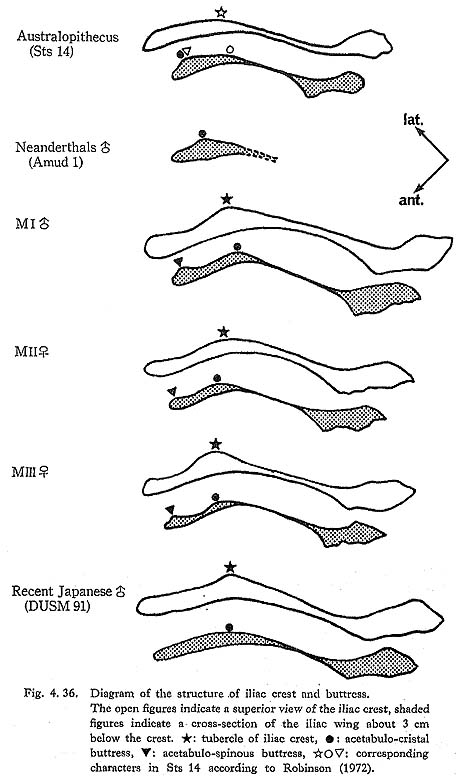
The shape of the upper part of the iliac wing (crest) of Minatogawa man appears as one-third of a geometrical circle, the arch of which is slightly depressed at the middle. In order to know the shape of the upper iliac wing metrically, the perpendicular measurement from the highest point of the iliac crest to the bispinal line (from sup. ant. spine to sup. post. spine), and distance from the superior anterior spine to the point of intersection of this perpendicular line (anterior part) are taken. The height and length of the anterior part are 63, 76 mm in MIl, 46, 63 mm in MIIr, 47, 70 mm in MIIIr, respectively. The ratio of the length of this anterior part to the bispinal length is 49% in MIl, 46% in MIIr, and 51% in MIIIr. This means that the highest point is located approximately at the middle of the wing. As far as the shape of the upper part of the ilium is concerned, the Minatogawa coxa is rather close to that of the Neanderthals but not to that of the Recent Sapiens (cf. Boule: 1911-13; McCown and Keith: 1939).
Iliac crest
The gradual sigmoid curvature of the crest from a superior view is rather weak, but the flexion at the tubercle of the crest and the anterior margin of the sacral part are sharp except in MII. The tubercle of the crest is situated more anteriorly than that of recent Japanese. Therefore, the ratio of the arch from the superior anterior spine to the tubercle to the whole arch from the superior anterior spine to the superior posterior spine is 27% in MIl and MIIr and 25% in MIIIr. The thickness of the crest at mid-point is small although thickness at the tubercle is not, except in MII.
Plate of the iliac wing
From the tubercle of the crest to the supra-acetabular region on the lateral surface there is a long prominent area (cf. Mednick, 1955). This is called the "vertical iliac bar" (McCown and Keith, 1939) or "acetabulo-cristal buttress" (Robinson, 1972) (Figs. 4. 34, 35). This buttress is marked in all Minatogawa ilia as it is in usual cases of Recent Sapiens. Anterior to this buttress there is another eminence from the superior anterior spine to the supra-acetabular region, which might be called the "acetabulo-spinous buttress" after Robinson (1972). This buttress is also seen in all the present specimens (Figs. 4. 34, 35).
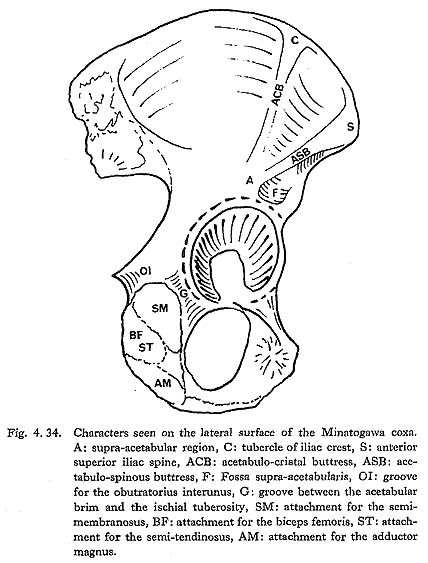
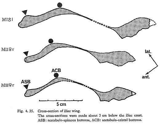
On the outer surface between the two buttresses a marked fossa is seen, and the corresponding part of the inner surface is slightly convex (Fig. 4. 35). The inferior anterior spine is well developed in all specimens. The notch between superior and inferior anterior spines (Incisura iliaca minor) is normal. Between the inferior spine and the acetabulo-spinous buttress, there is a supra-acetabular fossa in all specimens (Fig. 4. 34). The sulcus for the ilio-psoas muscles is well developed and rather deep. The fossa of the iliac wing is well developed in the medial surface of the wing; the central part of the outer surface is strongly concave. Therefore, the thickness of this region in all specimens is only around 1 mm. The gluteal line is marked in MI and MII but poor in MIII.
Sacral region
The iliac tuberosity is well developed in MIV, whereas it is poorly marked in MII and MIII. The condition in MI is not clear because of damage. The auricular facet is normal. On its margin the lippings of osteophytes can be seen but not marked. The preauricular sulcus (cf. Houghton, 1973) is well developed in all the Minatogawa coxa. The arcuate iliac buttress (arcuate bar, McCown and Keith, 1939) in the Minatogawa specimens is normal. Its thickness at the mid-point is 22 mm in MIr, 18 mm in MIIr, 17 mm in MIIIr and 16 mm in MIVr.
Acetabulum
The acetabulum is very small in the Minatogawa specimens. Maximum diameter is 48 mm on both sides of the MI, which is less than the mean for Japanese males and rather close to that of the females. Its depth is normal and the acetabular index is 52% in MIr and 50% in MIIr. No peculiar feature is seen on the acetabular brim of Minatogawa coxa.
Ischium
General view
The height of the ischium is fairly low (Tables 4. 21, 22), and the low height of the coxae is the result of this.
Greater sciatic notch
The notches of the Minatogawa females are normal, that is, the width and height are roughly equivalent to the mean of Japanese females. On the contrary, the notch of the male specimen (MI) is much wider and higher than those of the usual Japanese males. The height-width index of MI is normal, however. This notch width is partly due to the low position of the ischial spine.
Ischial tuberosity
It is well developed in all specimens having an ischium. In MIrl particularly, the area for the semimenbranosus and the area for the long head of the biceps femoris and the semitendinosus are wide with a ridge between them (Fig. 4. 34). This ridge is also seen in MII and MIII to a lesser degree. The width of the ischial tuberosity is 30 mm in MIl, 25 mm in MIIl and 23 mm in MIIIr. The tuberosity of the Minatogawa ischia is situated superiorly, that is, coming close to the acetabulum (Fig. 4. 36). The least distance between the acetabular brim and the tuberosity is 9, 8, 11, 9, 12 mm in MIr, MIl, MIIr, MIIl, and MIIIr, respectively. In this higher position, the groove for the obturator internus is located relatively low, that is, the upper margin of the groove is approximately at the same level as the upper margin of the tuberosity, especially in MIrl. In MIIrl and MIIIr the groove is situated slightly higher than the tuberosity (Fig. 4. 34).
Ramus of ischium
There are only two rami from MIl and MIIIr. The ramus of MIIIr is rather thin, while that of MIl is massive. No special characteristic is seen in either of these specimens.
Pubis
There is one complete pubis (MIII) and two incomplete pubes (MIr, MIIr).
General view
The length of the pubis is estimated to be 88 mm in MIr, which is somewhat larger than the mean for the Japanese males. It is 78 mm in MIIIr, less than that of Japanese females.
Upper ramus of pubis
The upper ramus seems stout in MIr and rather gracile in MIIIr. The shape of the cross-section is like a normal Recent Sapiens, that is, the sagittal diameter at the point where the outer and inner crests in the lower surface intersect is comparable to the transverse diameter at the same point.
Symphyseal region
The symphyseal surface of MIIIr is rather narrow. From the appearance of this surface, the age of this individual is estimated to be over forty according to the method of McKern and Stewart (1957). The fragments of the facets of MIr and MIIIr also show the age to be overforty. The pubic tubercle is well developed in MIIIr. The condition in the other pubes is not clear because of breakage. The area for the adductor longus and brevis is rough, forming a tubercle in MIIIr.
Lower ramus of pubis
The lower ramus is rather thick in MI?r but quite thin in MIIIr. No peculiar characteristic is found.
Sacrum
MI: male, only small pieces of auricular facets are preserved.
MII: female, lower left half is broken off, fifth lumbar vertebra is attaching on its upper surface. MIII: female, consists of the first segment, most of the auricular facets are preserved. MIV: female, consists of the first and second segments.
The maximum width is 100 mm in MII, 101 mm in MIII, 100 mm in MIV. These values are almost equal to the means for Neolithic and Recent Japanese males. The promontorium angle is 61°, 58°, 60° in MII, MIII, MIV, respectively.
Pelvis as a Whote
The pelvis was restored from the remaining coxae and sacra. The restored specimens show the height of the pelvis (coxa) to be small, as described before. Its width is estimated to be 232 mm in MII and 247 mm in MIII. These values are considerably less than the means for the female populations compared (Table 4. 23). It can be said that the Minatogawa female had a slender pelvis.
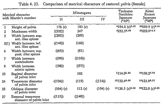
The transverse conjugate diameter of the pelvic inlet is estimated to be 106, 119, 114 mm in MII, MIII and MIV, and the oblique conjugate width is 104, 112, 104 mm in MII, MIII, MIV, respectively. These two values of MIII are normal, though somewhat smaller than the Japanese females, while the values for MII and MIV are extraordinarily small.
Discussion
Buttress of the iliac wing
It is well known that the acetabulo-cristal buttress and the acetabulo-spinous buttress are seen in humans and nonhuman primates, respectively. In the course of evolution, the acetabulo-cristal buttress changes its position from anterior to posterior, for example, it is located at a point about one-eighth of the iliac width posterior from the superior anterior iliac spine in the Australopithecus, at a point of about one-fifth in the Neanderthals, and about one-third in the Recent Sapiens (Fig. 4. 36). The buttress of the Minatogawa ilium is located at a point about one-fourth from the anterior end. This anterior position is sometimes seen in the Neolithic and Recent Japanese.
The presence of the acetabulo-spinous buttress is quite rare in human ilia. There are a few cases to have it in the Neolithic (Jomon) Japanese, though it is weakly developed. According to Robinson (1972), a strong acetabulo-spinous buttress and a weak acetabulo-cristal buttress are seen on the ilium of Sts 14 (Fig. 4. 36), but it is doubtful that they are homologous to those of Recent Sapiens. The functional meaning of co-existence of the two buttresses should be interpreted by Roux's law "The bone is so made as to obtain the maximum strength by means of the minimum amount of material"-that is, the force acting on the superior anterior iliac spine by the muscles and ligaments may be strong relative to the amount of bone mass. Accordingly, the abdominal muscles and tensor fascia latae may be relatively strong in Minatogawa people.
Superior position of the ischial tvherosity
It is well known that the ischial tuberosities, including the obturator groove, are upwardly displaced in the Neanderthals, and that most of the Mt. Carmel men show in this respect an intermediate condition between the Neanderthals and Recent Sapiens. The Minatogawa ischium also indicates an intermediate condition. This condition is often seen in the Neolithic Japanese and sometimes in Recent Japanese to a lesser degree. The functional meaning of this upward position is not clear, but the fact that the attaching muscles incline supero-anteriorly when the hip joint is fully flexed suggests that it may be due to a flexed position such as squatting. This is reinforced by the fact that this type of ischial tuberosity is often seen in such habitual squatters as Indians.