CHAPTER 4
Postcranial Skeleton of the Minatogawa Man
Hisao Baba* and Banri Endo**
*Department of Anatomy, Dokkyo University School of Medicine;
**Department of Anthropology, Faculty of Science,
The University of Tokyo
| ( 2 / 7 ) |
|


  |

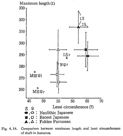
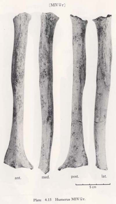
Except in MIV the circumference at the mid-shaft in Minatogawa specimens is equivalent to that of Luzon Negrito (Genet-Varcin, 1951), and is very low compared with that of other populations (Fig. 4. 15).
The torsion angle is considerably smaller than those of other populations compared, except in the case of Neolithic Japanese where it is slightly larger.
Upper Epiphysis
Articular head
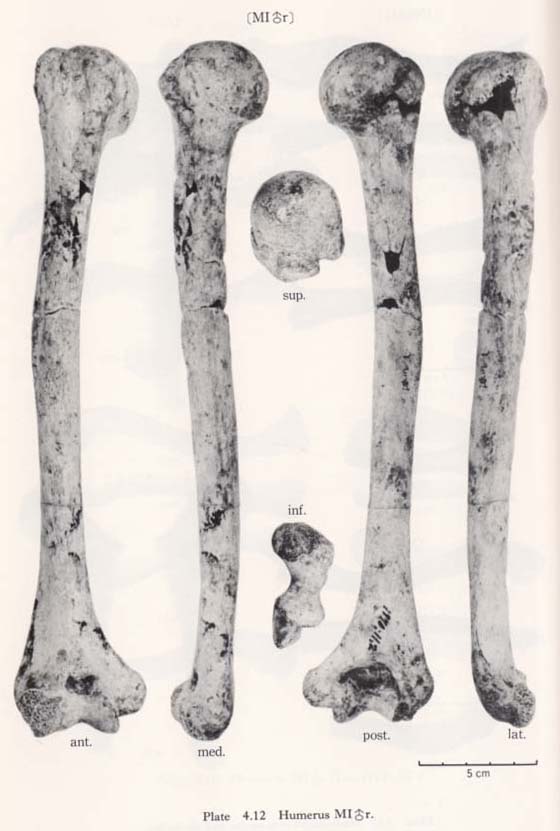
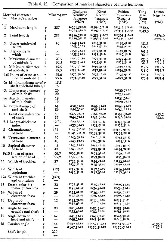
The angle between head and shaft varies from 40° to 44° in Minatogawa humeri. These values come within the range of the Neolithic Japanese, but they seem less than that of the Recent Japanese. When the head is medially viewed, its long axis is slightly inclined from the longitudinal axis of the upper shaft (postero-superior to antero-in-ferior).
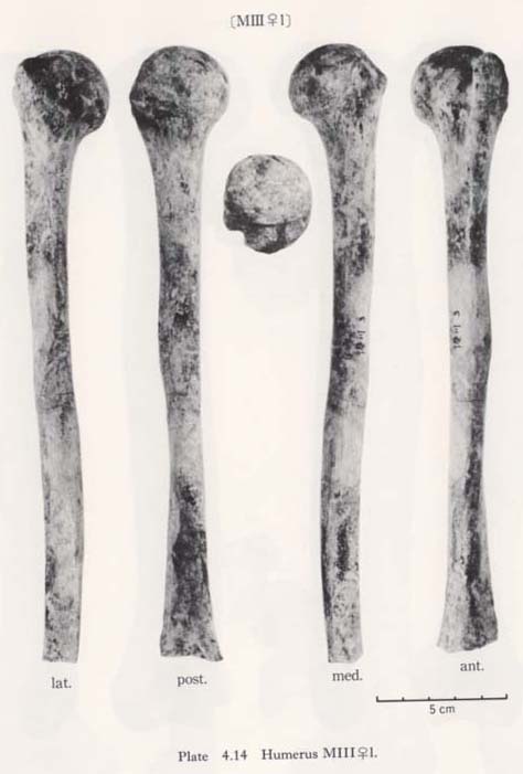
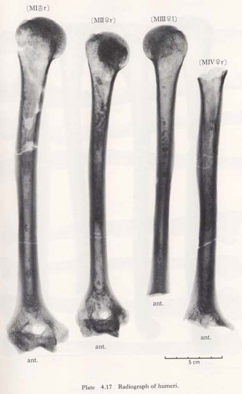
The size of the articular head is slightly smaller than the means of the compared populations (Tables 4. 12, 13). In the case of Minatogawa humeri the vertical diameter is much larger than the transverse as compared with other populations. The articular surface and margin are somewhat rough and a hyperostotic feature is seen in MI r.
r.
Greater tubercle
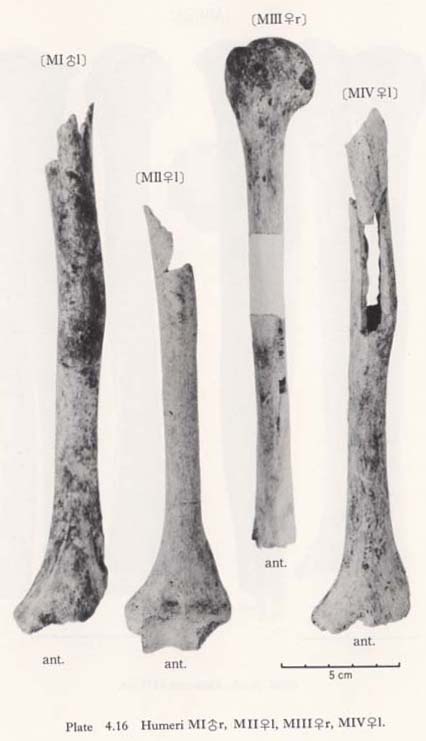
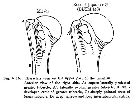
This tubercle is more or less developed and projects supero-laterally forming a right angled corner on the upper end in all the Minatogawa specimens (Fig. 4. 16-A). Its superior surface is located approximately at the same level as the articular head. From an anterior view its lateral surface shows a straight line having no swelling as in the Recent Japanese (Fig. 4. 16-A'). Areas for the supraspinous, infraspinous and teres minor muscles are well defined. Hyperostotic lippings are seen on the supero-lateral border of the tubercle in MIIr.
Lesser tubercle
Its development is normal and the area for the subscapularis muscle is seen on its upper surface.
Intertubercular sulcus
It is deep but not wide in Minatogawa humeri forming a half circle in cross-section (Fig. 4. 16-D). Its diameter is 5.5 mm in MIr, 5 mm in MIIr and MIIIl. It seems that they had thin tendons of the long head of the biceps brachii muscle.
Diaphysis
General character
The most striking character of the Minatogawa humeral shaft is the antero-lateral flexion around mid-shaft. This type of flexion cannot be seen in the Recent Japanese. It is, however, sometimes seen to a lesser degree in the robust Neolithic Japanese.
Its diameter is also characteristic, i.e. very thin in relation not only to the total length but also to the diameter of the ends (Tables 4. 12, 13).
Muscle relief
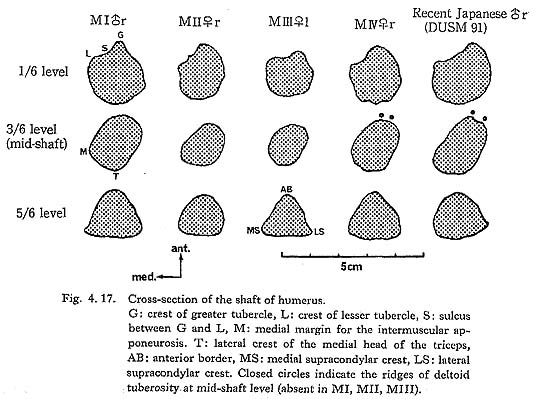
In all the Minatogawa humeri, the crest of greater tubercle is well developed, which implies the presence of a strong pectoralis major muscle (Figs. 4. 16-B, 17-G). Par ticularly in the male, it becomes a sharp prominent ridge and continues to the medial crest of the deltoid tuberosity. When the usual humerus with straight shaft is put hori zontally on its dorsal surface, the contour of this crest inclines from proximal to distal. In Minatogawa humeri, on the contrary, it is horizontal.
The deltoid tuberosity is also remarkable in the male. In MIIr it is well defined but rather poor. In MIIIl its lateral crest is well developed. Generally the deltoid muscle of the Minatogawa man is presumed to be relatively strong.
The crest of the lesser tubercle is also well developed in MIrl, MIIIrl, MIVrl, while it is faint in MIIr. Accordingly, the intertubercular sulcus expands downward between the two crests, except in MIIr (Figs. 4. 16, 17-S).
The medial margin for the intermuscular apponeurosis (Fig. 4. 17-M) is generally sharp and extends upward. It directly continues to the ridge for the medial head of the triceps brachii muscle. The lower part of the anterior border (AB) is sharp and prominent in MIIIrl, somewhat sharp in MIrl, MIVrl, and round in MIIrl.
On the dorsal surface, the sulcus for the radial nerve is well defined in Minatogawa humeri except in MIIrl. The upper part of the lateral crest for the medial head of the triceps runs vertically in the Minatogawa humeri, demarcating the dorsal border of this sulcus. This crest is remarkable in MI and MIV but rather poor in MII and MIII.
The dorsal convexity at the lower part of the shaft is pronounced in MIVrl, moderate in MIrl and MIIIrl, and slight in MIIrl.
Cross-section
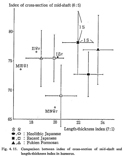
The shape of the cross-section of the mid-shaft is somewhat flattened in the Minato gawa specimens but is not as flat as in the usual cases of Japanese (Figs. 4. 15, 17), The cross-section at the deltoid tuberosity is rectangular in MI and MIV through the development of four crests, namely, two deltoids, the medial margin (M) and one for the lateral crest of the medial head of the triceps (T).
Lower Epiphysis
Articular part
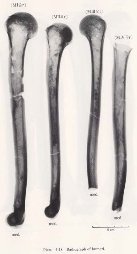
The trochlea and capitulum are moderate in size in the male (MIr) and the female (MIIrl). The groove of the trochlea of MIr is so deep that the dorso-ventral diameter at the groove becomes 13 mm, only 57% of that at the medial end. This value of 13 mm is even less than that in the female (14 mm in MIIr).
Other characters
The medial epicondyle is well projected in all the specimens. The area for the flexor muscles of the forearm is well denned. The olecranon fossa is large in MIr and moder ate in MIIrl, In MIVl the supratrochlear foramen is present. The radial and coro noid fossae are normal. In MIIrl the hyperostotic change is seen in these three fossae.
The condylo-diaphyseal angle of the Minatogawa specimens shows high values as compared with the Japanese and the Formosan. This implies that the elbow of the Minatogawa man was straight.
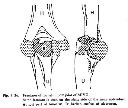
The fracture of the distal end in MIVrl
The condyle is broken off in MIVr and 1 as above mentioned. There is a peculiar feature noted in these fractures: their shape and position are exactly alike (Plates 4. 15, 16). Some parts of the fractured surface are covered by carbonate. These fractures will be discussed with the fracture on the olecranon.
Discussion
Flexion at mid-shaft
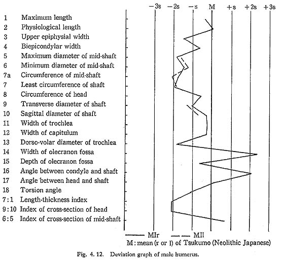
Generally, some humeri having the well-developed deltoid tuberosity seem to possess strong flexion at mid-shaft. However, the upper shaft is always parallel to the lower shaft in these cases. On the contrary, the axis of the upper part of the shaft makes an angle of about 8° to the axis of the lower part in MIr. This angle is 6° in MIVr and 7° in MIIr and MIIIl.
As mentioned, this type of flexion is seldom seen in the Neolithic and Recent Japanese, but sometimes seen in the Neanderthals as La Ferrassie (McCown and Keith, 1939). It is also seen in the Firelander humerus cited in Martin's textbook (1928).
Slenderness of the shaft
The humeral shaft in Minatogawa man is generally very thin, but it shows marked development of the muscle relief. In usual cases of the Neolithic and Recent Japanese, the relief of the muscle is almost invariably proportional to the robustness. In other words, a thick bone has well-developed muscle relief.
In this respect, Minatogawa humeri bear close resemblance to that of the Firelander cited in Martin's textbook, and some resemblance to the Sinanthropus humeri (Wei denreich, 1941). In addition, they completely differ from the robust Neanderthal humeri.
Medial inclination of the lower articulation
As Fig. 4. 18 shows, when the axis of the articular head (A) of MIr is positioned parallel with those of the Neolithic and Recent males, the axis of the lower articular surface (C) of MIr faces more medially than the others. The angle between the axes of head and lower articulation reaches 56° in MIr, whereas it is 35.5° in the Neolithic Japanese and 40.5° in the Recent Japanese.
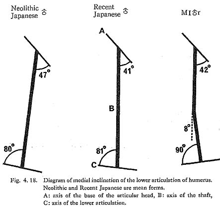
There is no reasonable interpretation for the functional meaning of this medial incli nation. It seems, however, that the shortness of the clavicle in Minatogawa man might have some relationship to it, because if the axis of the lower articulation of the MIr is set in the same way as the comparative humeri, the head of MIr will be situated more medially than the others.
ULNA
Materials
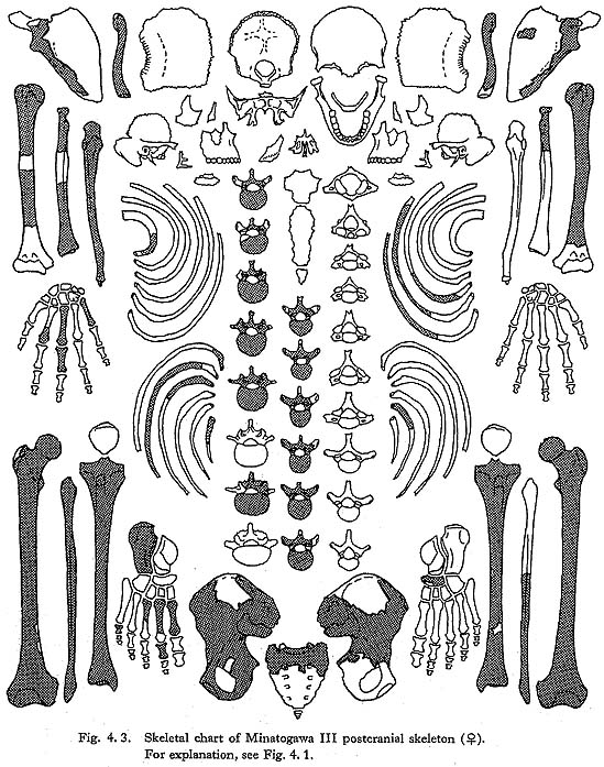
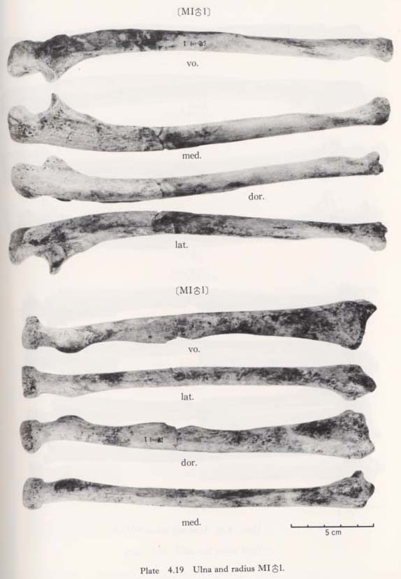
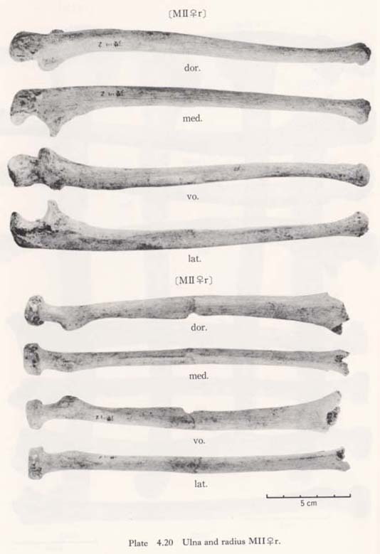
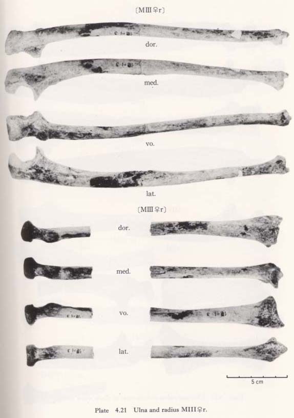
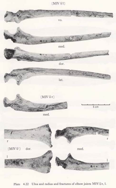
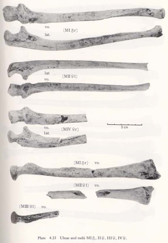
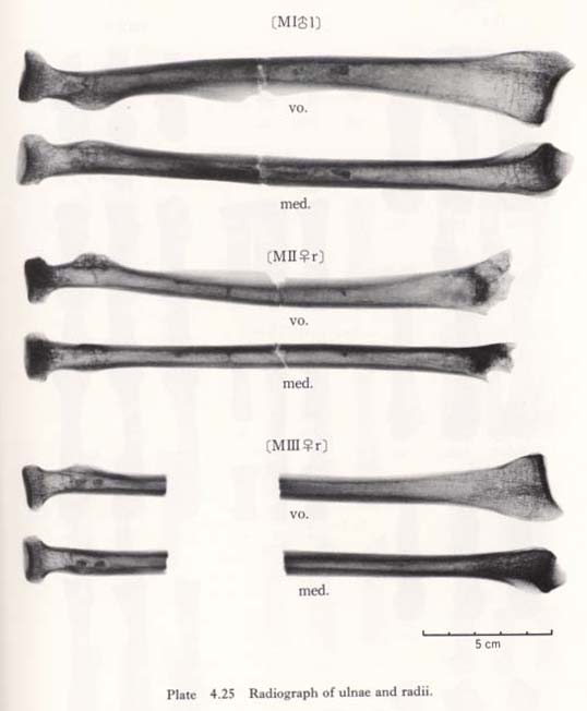
There are seven ulnae, belonging to four individuals (Figs. 4. 1-4, Plates 4. 19-25).
MIr: male, complete.
MII: male, complete.
MIIr: female, nearly complete, styloid process broken off.
MIII: female, nearly complete but distal end broken off.
MIIIr: female, complete.
MIVr: male-like female, upper half.
MIVl: male-like female, nearly complete but distal end broken off.
General Character
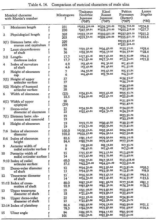
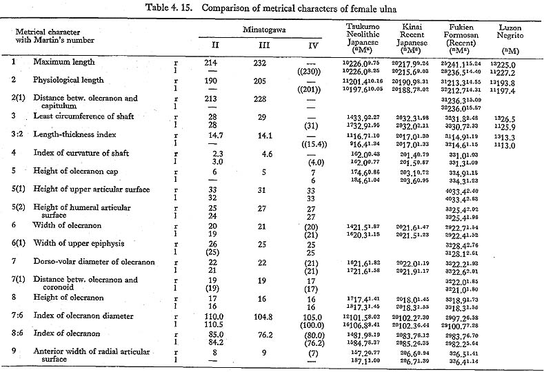
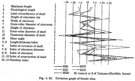
MI and MII ulnae are considerably shorter than the ulnae of the other populations, while MIII and MIV are longer than the others except the Fukien Formosan (Figs. 4. 19, 20; Tables 4. 14, 15). MI is as robust as those of the Japanese and more robust than those of the Fukien Formosan and Luzon Negrito. The Minatogawa females had rather gracile ulnae like the Fukien Formosan, but they are not as gracile as those of the Luzon Negrito.
  |
Upper Epiphysis
Articular surface
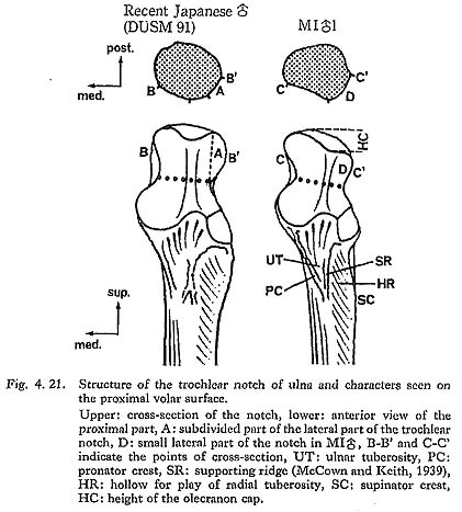
The trochlear notch of the Minatogawa ulna is quite different from that of the Recent Japanese. In usual cases, the central ridge divides the notch cavity into lateral and medial halves, and the lateral half is slightly smaller than the medial (Fig. 4. 21). The lateral half is again divided into two parts. The lateral one-third of its proximal part, which contacts the lateral wall of the humeral trochlea in case of extension, is distin guishable (Fig, 4. 21-A). On the contrary, the lateral half in Minatogawa ulna is very small and situated distally (D). In addition, it faces laterally having no clear subdivision. The radial notch is normal.
Olecranon
Its overall size is normal. Both the depth and the height are slightly smaller than the means for the Japanese or Fukien Formosan. From a lateral view, the olecranon seems very high (Fig. 4. 21-HC). However, this is because of the distal location of the proximal border of the lateral part of the trochlear notch. Accordingly, the height of the olecranon cap becomes very large in the Minatogawa specimens. A peculiar breakage is seen in the olecranon of MIV where the dorsal parts of both right and left olecranons are broken off.
Coracoid process
Generally the coracoid process is well projected anteriorly but is rather thin, because its distal surface is depressed. The ratio between the depth of the coracoid and that of the olecranon indicates the development of the coracoid; in other words, the degree of upward orientation of the trochlear notch is marked. This ratio varies from 132 to 148 in Minatogawa ulnae, showing Recent Sapiens character.
Diaphysis
General character
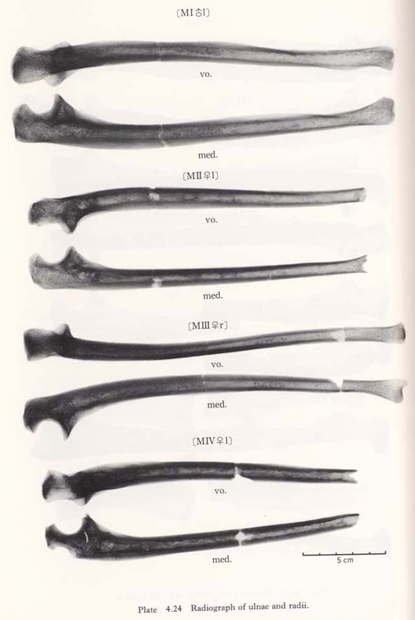
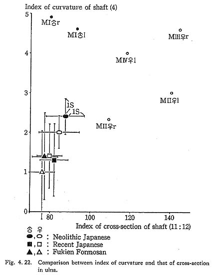
The most impressive feature of the shaft in Minatogawa specimens is the pronounced dorsal curvature. The index of curvature is 4.9 in MIr, 2.3 in MIIr, 4.6 in MIIIr, and 4.0 in MIVl. Except in MIIr, these values are about twice as large as the mean for the Neolithic Japanese and more than three times larger than that for the Recent Japanese or Fukien Formosan (Fig. 4. 22), In this point Minatogawa ulnae exceed most of the fossil ulnae, as far as we know, except Skhül VII, whose value is 5.2. Increase of the dorso-volar diameter of the upper part of the shaft to the coronoid process is very slight and resembles that of the Neanderthals (Endo and Kimura, 1970).
Surface structure
The pronator crest (Figs. 4. 21-PC, 23-PC) is well developed to form a sharp edge extending to the anterior margin in all the Minatogawa specimens except MIIIr, The supinator crest (SP) is also marked. It continues to the interosseus margin except in MIrl, in which it extends onto the lateral surface. There is another crest (supporting ridge, SR, McCown and Keith, 1939) running from the medial margin of the radial notch to the anterior margin. It is prominent in all these specimens, as seen on the cross-sections of Fig. 4. 23.
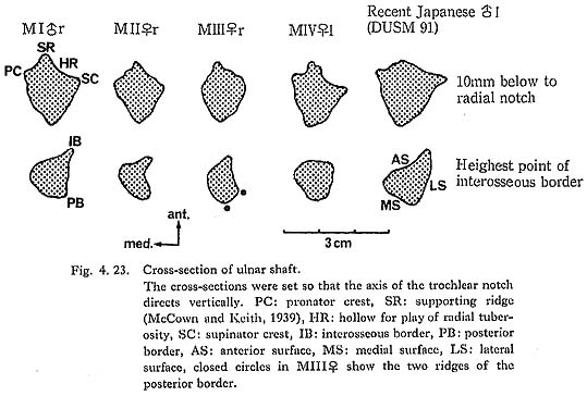
Between this ridge and the pronator crest, the ulnar tuberosity (UT) is seen in the shape of a triangular pit. In MIIIr, this ridge expands downward along the axis of the upper shaft, then turning laterally it reaches the interosseus margin. The hollow for play of the radial tuberosity (HR) is marked in MIrl and MIVrl, slight in MIIrl, and absent in MIIIr.
The dorsal margin is sharp in MIrl, MIIrl and MIVrl, while in MIIIr it is not only blunt but flat, having two edges with distances of 5 mm at the mid-point. As usual, the area for the anconeus is seen on the medial surface of the proximal part.
The interosseus margin is prominent in MIrl, but is rather poor in the rest of the specimens (Fig. 4. 22). Particularly in MIIIr it is very short, being similar to those of the Neanderthals. The ridge for the pronator quadratus is strongly developed having a round edge in MIrl, but it is poor in the other specimens.
Cross-section
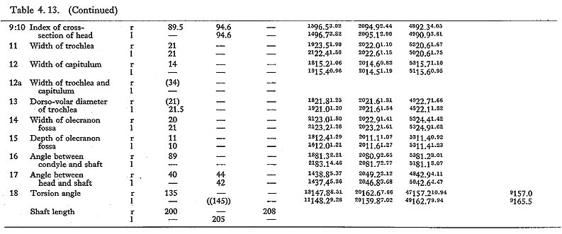
The shape of the cross-section at the lower end of the radial notch is moderately flattened in Minatogawa ulnae. That is, the index of platoleny varies from 78.3 to 89.5. This range is slightly larger than that for the Recent Japanese, but within the range of the Neolithic Japanese or Fukien Formosan, and considerably larger than that of the Luzon Negrito. In addition, it is far smaller than those for the Neanderthals. As for the cross-section at the mid-shaft, Minatogawa ulnae, except in MIrl, show transverse flatness due to the anterior location of the interosseus margin (Fig. 4. 23-IB), The ratio of the diameters at the mid-shaft falls within the range of 109 to 144 in the females (Fig. 4. 22). This range is extraordinarily large as compared with the female populations, but this value of MIrl falls in the normal range.
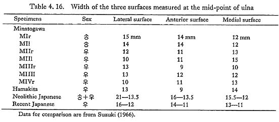
To clarify the shape of the cross-section at this point, we measured the widths of the three surfaces on the cross-section according to Suzuki (1966). As Table 4. 16 shows, the width of the anterior surface in the Minatogawa female ulnae is generally small, and the width of the medial surface is larger than those of the other two (MIIrl and MIVl). This condition is similar to that of the Hamakita ulna, which is regarded as the late Pleistocene man in central Japan (Suzuki, 1966).
Distal Epiphysis
The transverse diameter of the capitulum is 15 mm in MIrl, 14 mm in MIIrl and MIIIr. The styloid processus is normal. No other peculiar characteristics are seen.
Discussion and Conclusion
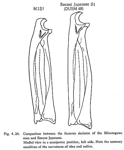
Dorsal curvature of the shaft
As mentioned above, Minatogawa ulnae have strong dorsal curvature (Figs. 4. 22, 24). In this point they resemble those of the  VII or the chimpanzee. But the curvature in Minatogawa ulnae is present only in its proximal part, while in VII and the chimpanzee it extends over all the shaft. Accordingly, this type of curvature in Minatogawa ulnae seems unique.
Olecranon cap
Since the lateral half of the trochlear notch is small and situated distally, the olecranon cap seems to be high in Minatogawa ulnae, as above mentioned (Fig. 4. 21-HC). This high type of olecranon cap is also seen in most of the Neanderthals. However, the height of the cap in the Neanderthals is due not only to the peculiar shape of the trochlear notch but also to the upward projection of the olecranon itself. In Minatogawa ulnae, the height of the olecranon is normal, though its end comes downward. Thus it seems that there is no functional difference of the elbow extension between Minatogawa man and recent man.
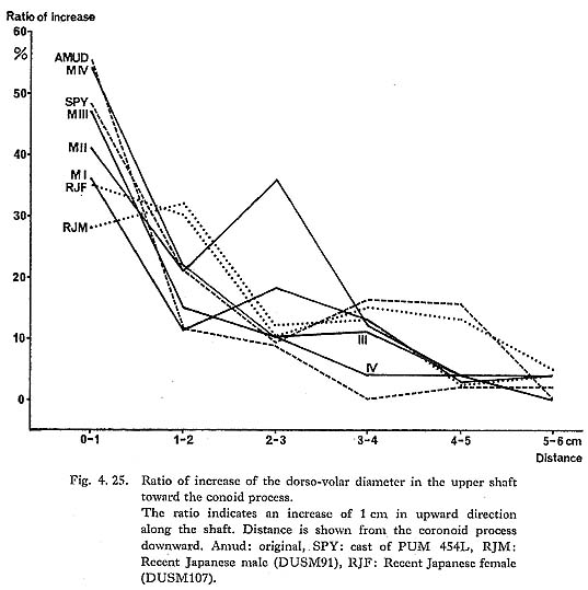
Dorso-volar diameter of the upper shaft
It is well known that the sudden increase of the dorso-volar diameter of the upper part of the shaft below the coronoid process is a significant characteristic of the Nean derthal ulna (McCown and Keith, 1939). This characteristic indicates a striking contrast to the gradual increase in the Recent Sapinens. For comparison we measured the dorso volar diameters at intervals of 1 cm from the level of the coronoid process accrding to Endo and Kimura (1970). As Fig. 4. 25 shows, Minatogawa ulnae have a marked increase near the coronoid process, indicating close resemblance to those of the Neanderthals and differing from those of the Recent Japanese as well as Europeans.
Fractures in MIV
When the humerus and the ulna are Jointed at the elbow and viewed from a dorsal direction, the lost part of the humerus (Fig. 4. 26-A) overlaps the fracture of the olecranon (B). This condition occurs on both sides in similar shapes. Hence, the cause of the formation of these fractures might not have been accidental.