CHAPTER VIII MICROSCOPIC STUDY ON THE "AMORPHOUS SILICA" IN SEDIMENTS FROM THE DOUARA CAVE
Akiko MATSUTANI
Department of Cultural Anthropology, The University Museum, The University of Tokyo
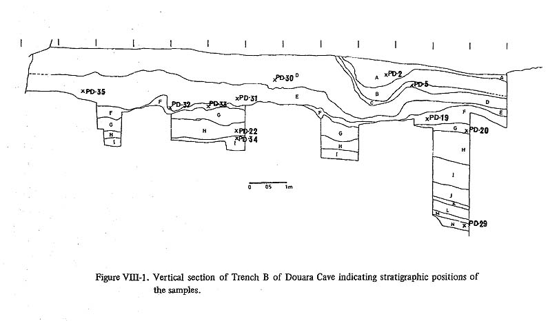
1. INTRODUCTIONAs described in Chapter VI, some sediments from the Douara Cave contained "amorphous silica". Under the microscope these reveal structures that resemble those in plant epidermis, and it was suspected that these might be opaline silica or silica skeletons of certain plants. The author was asked to study these and if possible to identify the plant species from which they derive. 2. MATERIALS AND METHODSEleven cave samples, ash from the fireplace in layer E, and one surface and one bedrock sample were treated (Table VIII-1, Fig. VIII-1). In addition, concretions adhering to flint implements (Layers E and F) were also treated.
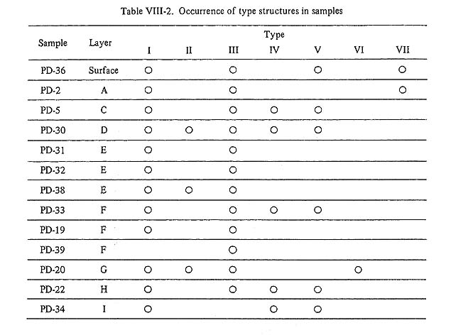
The following procedures were used. First, two or three grams of each sample were poured into a beaker, and diluted hydrochloric acid (10%) was added until no more foam appeared. Then thie sample was washed with distilled water three or more times, desiccated in an oven and mounted in Caedax. These preparations were observed under a light microscope and photographed at magnifications of × 200 or × 400. Some samples (PD-19, PD-34, ash from the fireplace) were also treated with a solution of tribromoethane and benzene at a specific gravity of 2.1. But there was not marked difference between the preparations treated in this way and those treated with only hydrochloric acid. 3. RESULTS AND DISCUSSIONVarious structures were extracted from most samples except those from the lowest layer, the ash from the fireplace and the bedrock (Table VIII-2), Some samples contained many structures, others only a few. These structures were found most abundantly in the samples from Layers F and G, but a few were found even in the concretions adhering to stone implements from Layers E and F. These structures can be grouped into seven types.
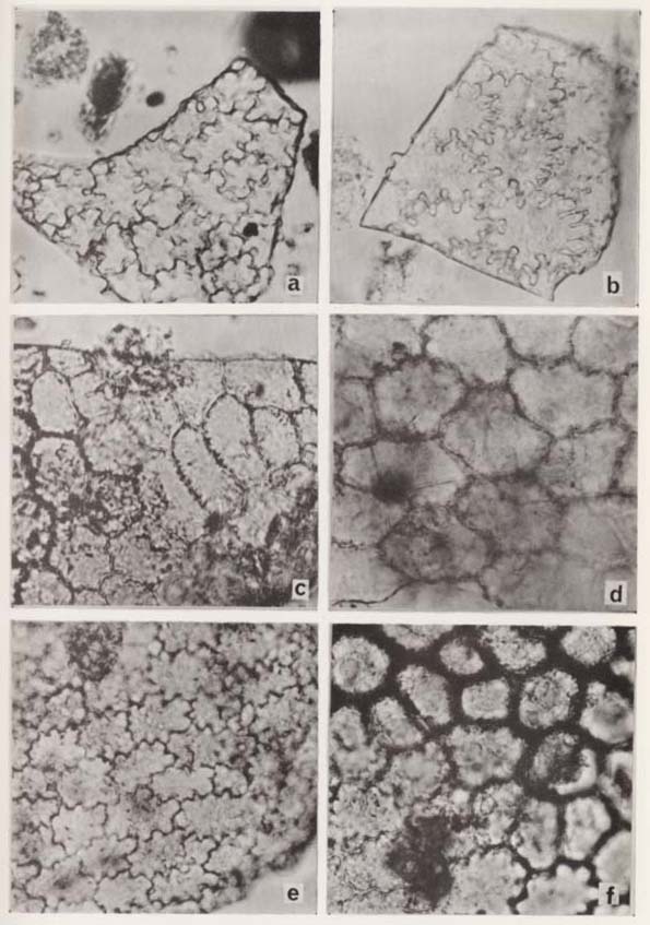
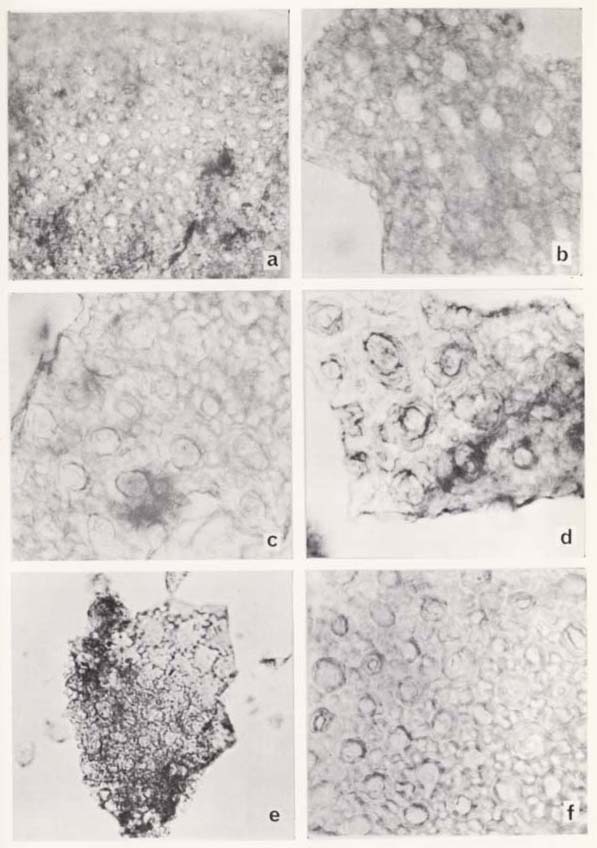
Type I structures (Pl. VIII-1) were observed most abundantly and in most samples. They greatly resemble plant epidermis; their shapes vary between polygonal and strongly wavy. However, these forms are observed in such a wide range of plants, and they cannot be used as identification keys.
Type II structures (Pl. VIII-2) resemble Type I, but they have a small circular knob in their centers. This type was found in sample PD-30 (Layer D), in the concretions adhering to a flint implement from Layer E, and most abundantly in sample PD-20.
Type III structures (Pl. VIII-3) were also extracted from most of the samples, except those from Layers I and N, the ash from the fireplace and the bedrock. They seem to have a thick wall around the cell hollow, and some also have a small circular knob in their center. As seen in Plate VIII-3 c, d, this type is linked with Type I or Type II.
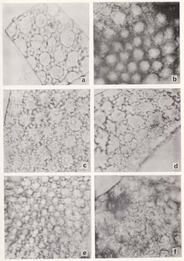
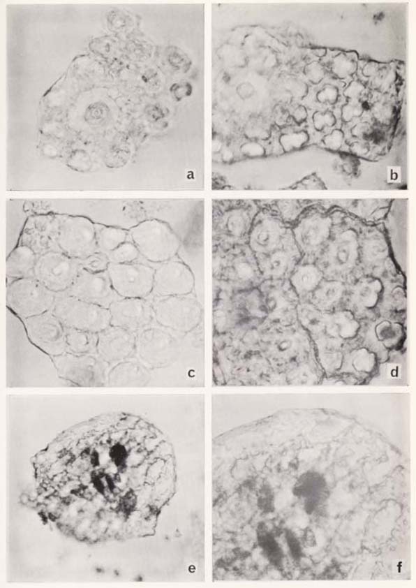
Type IV structures (Pl. VIII-4) occurred in samples PD-5, PD-30, PD-33, PD-22 and PD-34. In this type, smaller or larger hollows were observed over the surface, and in some of them (Pl. VIII-4c, d) protuberances around the hollow were observed. Type III and Type IV seem to occur together (Pl. VIII-4e).
The structure seen in Plate VIII-4 f was extracted from sample PD-20. This structure has a protuberance around the small and large hollows, and the areas around these are filled by small rectangular shapes. Type V structures (Pl. VIII-5a, b) were found in several samples as shown in Table 2. Type VI structures (Pl, VIII-5c, d) were extracted only from sample PD-20. Type VII structures (Pl. VIII-5e, f) were found only in sample PD-2. Small circular and oval shapes occur in Types V and VI.
All types except Type I seem to have a common structure in that they have small circular or oval knobs in their centers. Further, as seen in some photographs, some of these types occur together. It is probable that these come from the tissues of one plant. However, it cannot be firmly concluded that these are the silica skeletons of plant tissues until they are matched to living plants. It is well reported that Gramineae, Cyperaceae, and Equisetum deposit abundant silica in the epidermis and that silica skeletons adhering to archaeological materials can be used to identify the plants utilized by ancient men (Schellenberg, H. C., 1908; Edman, G. and E. Söderberg, 1929; Helbaek, H., 1959, 1961; Watanabe, N., 1968, 1970; Matsutani, A., 1972). Investigation of plant Opal or opaline silica has proved helpful in the reconstruction of past floral environments (Kanno, I.and S.Arimura,1958: Smithson, F. 1958; Rovner, I., 1971). However, the structures reported in these studies were not observed in the samples from the Douara Cave. Thus it was necessary to make comparisons with the silica structures of living plants. Spodographic preparations were made by incinerating one square centimeter of a leaf or other part of a living plant in a porcelain dish over a Bunsen burner. Spodograms of some Gramineae, Cyperaceae and Equisetum were available, but the Douara samples do not resemble them. Moreover, the Douara samples do not suggest a monocotyledonous origin. According to F. Netolizky (1912) many dicotyledons also reveal silica skeletons. Spodograms were made for some dicotyledons such as Salsola sp. (Chenopodiaceae),Amalasis syriaca (Chenopodiaceae), Peganum harmala (Zygophyllaceae), Aellenia arteani (Chenopodiaceae), Cyperus papyrus (Cyperaceae), Avena sp. (Gramineae) collected by Prof. I. Kobori from around Douara Cave. When dealing with remains from human living sites, seeds or fruits also must be considered, so spodograms were made for such seeds and fruits as pistachios and dates. Other plants including some ferns and mosses were also submitted to the study. In all cases the comparisons proved non-committal or negative, leaving the silica structures from Douara unidentified for the present. BIBLIOGRAPHY
|