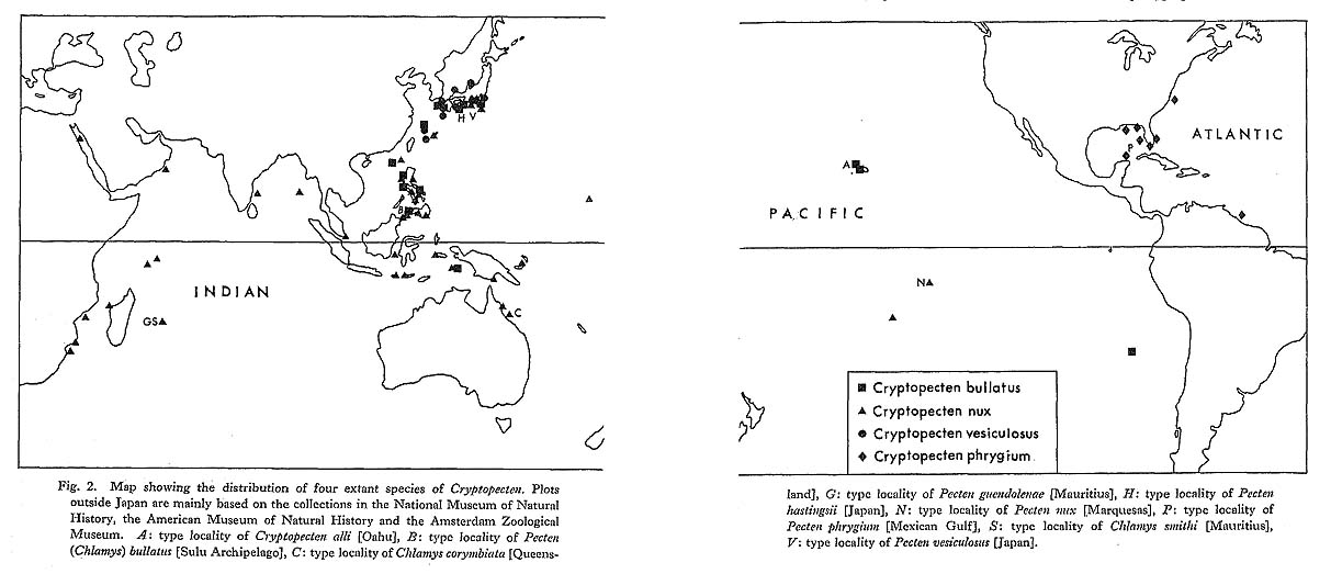
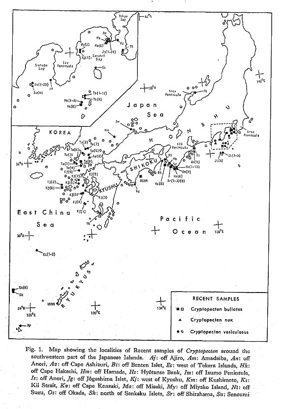
CHAPTER 5
Ontogenetical Development of Surface Sculpture in Cryptopecten
|
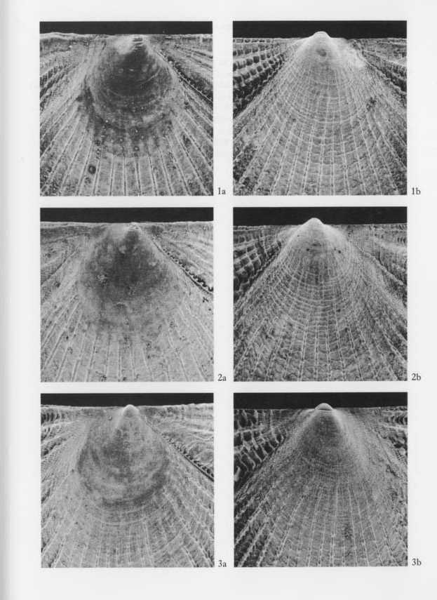
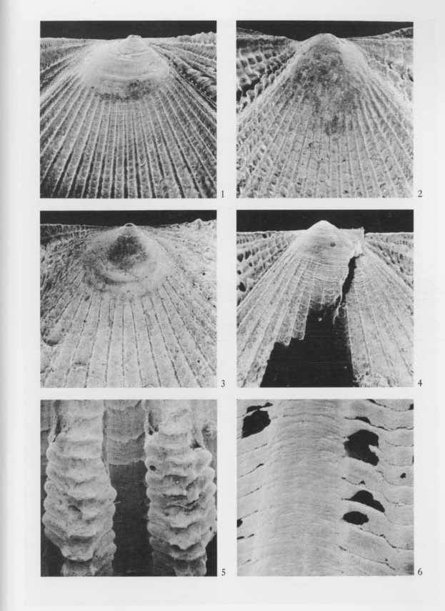
The surface sculpture of Cryptopecten is unique and clearly different from that of other pectinid genera. Because the present evolutionary study depends to a great extent upon the variability of radial ornamentation, it will be necessary to clarify the detailed structure and ontogenetical development of surface morphology in each representative species. The surface morphology of the following nine selected individuals was examined by use of a scanning electron microscope (Hitachi: S-700) with relatively low magnification. Specimen 1 (UMUT RM16002a). Conjoined (living) valves of Cryptopecten bullatus, belonging to Sample Hs (3) (B). 13.2 mm long, 12.4 mm high, 3.8 mm thick. [Plate 10, Fig. 3; Plate 11, Fig. 3]
Specimen 2 (UMUT RM16059a). Conjoined (living) valves of Cryptopecten vesiculosus [Pheno- type R]*, belonging to Sample Hy. 10.6 mm long, 11.0 mm high, 4.4 mm thick. [Plate 10, Fig. 2; Plate 11, Fig. 2]
Specimen 3 (UMUT RM16059b). Conjoined (living) valves of the same species [Phenotype Q]*, belonging to the same sample. 11.6 mm long, 11.8 mm high, 5.0 mm thick. [Plate 10, Fig. 1; Plate 11, Fig. 1]
Specimen 4 (UMUT RM16061a). Conjoined (living) valves of the same species [Phenotype R], belonging to Sample Jg (2) (for the observation of transverse section and ventral margin of adult shell). 25.3 mm long, 25.6 mm high, 11.8 mm thick. [Plate 12, Fig. 6; Plate 13, Fig, 2]
Specimen 5 (UMUT RM16138a). Conjoined (living) valves of the same species [Phenotype Q], belonging to Sample Is (for the observation of transverse section and ventral margin of adult shell). 27.3 mm long, 25.4 mm high, 12.7 mm thick. [Plate 13, Fig. 1] Specimen 6 (UMUT CM16015a). Right (fossil) valve of Cryptopecten nux, belonging to Sample Kk (N). 12.2 mm long, 12.1 mm high, 3.5 mm thick, [Plate 12, Fig. 1] Specimen 7 (UMUT CM16015b). Left (fossil) valve of the same species, belonging to the same sample. 12.0 mm long, 12.1 mm high, 2.7 mm thick, [Plate 12, Fig. 2] Specimen 8 (UMUT CM16170a). Right (fossil) valve of Cryptopecten spinosus sp. nov., belonging to Sample Kk (S). 12.5 mm long, 12.3 mm high, 3.0 mm thick. [Plate 12, Figs. 3, 5]
Specimen 9 (UMUT CM16170b). Left (fossil) valve of the same species, belonging to the same sample. 12.9 mm long, 13.2 mm high, 3.0 mm thick. [Plate 12, Fig. 4]
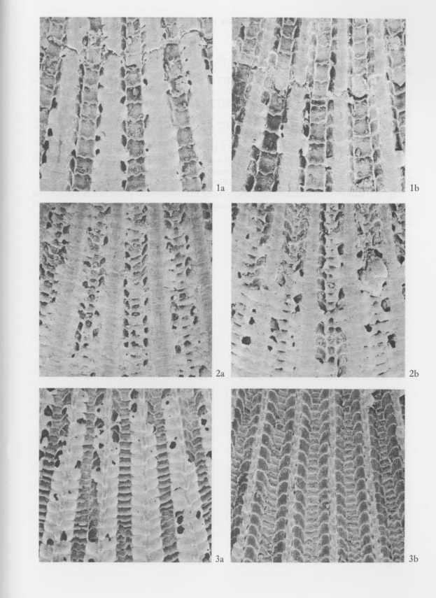
Because the umbonal area of full-grown shells is commonly abraded or concealed by encrusting organisms, suitable specimens for the observation of early stages are restricted to young individuals such as those of specimens 1-3, 6-9. As the young shells of C. bullatus, C. vesiculosus (both phenotypes), C. nux and C. spinosus share many common surface characteristics, the ontogenetical development of surface sculpture is collectively described below. Prodissoconch.-The prodissoconch is well demarcated on the umbonal surface in these young individuals (Specimens 1-3, 6-9). It is D-shaped in outline without wings, strongly inflated in both valves and as large as 140-200 microns in length. The surface is apparently smooth. C. bullatus seems to have a slightly larger prodissoconch than three other species. As is commonly the case in bivalves (e.g., Ockelmann, 1956; Waller, 1981), the prodissoconch is generally composed of two parts: a smooth or radially striated part near the origin of growth (prodissoconch I) and a commarginally striate part (prodissoconch II). The two parts are not clearly distinguishable in these examined specimens, but this may be due to subsequent abrasion, [See Plates 10 and 12] First stage of dissoconch (shell height less than 1.5 mm).-The shell of this stage is strikingly inequivalve and inequilateral, left-convex and pteriform with indistinctly demarcated posterior wing. The outline reminds one of some aviculopectinids from the Upper Paleozoic and Lower Mesozoic. The byssal notch becomes gradually distinct, but a ctenolium is still undeveloped. The disk and wings of the right valve are smooth except for weak growth-lines. In contrast, the left valve is ornamented with numerous Campto-nectes-like divaricate striae which are also distributed on both wings. In the later half of this stage radial ribs appear on the disk of the left valve, though their number and strength are still unstable owing to irregular insertion, convergence and disappearance. [See Plates 10 and 12] Second stage of dissoconch (shell height between 1.5 mm and 4 mm).-In this stage the outline of shell changes from pteriform to pectiniform and from left-convex to equi-convex. The posterior wing becomes well delimited, and several denticles of the ctenolium are developed along the anterodorsal margin of disk. Camptonectes-like striae are persistent on the disk and wings of the left valve and appear also on the disk and posterior wing of the right valve. Radial ribs develop on the disk of the right as well as the left valve. They are sometimes flexuous but simple, provided with fine scales, neither bifurcated nor inserted, and always much narrower than the interspaces. Their number varies with the species (in the examined specimens, 22 in C. bullatus, 16 or 17 in C. vesiciilosus and 22 in C. nux). A few much broader radial ribs appear on the wings of both valves, and on the left anterior wing they form a striking lattice sculpture with strong concentric lamellae. [See Plates 10 and 12] Third stage of dissoconch (shell height more than 4 mm).-The shell is nearly acline and equiconvex in the earlier half of this stage but becomes right-convex and slightly pro-socline in the later. Denticles of the ctenolium continue to develop throughout this stage. The Camptonectes-like striae disappear entirely by the beginning of this stage. Radial ribs are persistent except for one or two on the peripheral areas of the disk, relatively narrow at first but apparently much broadened later. The broadened radial ribs show a very unique structure. As clarified by SEM, each broadened rib consists of a central solid ridge and a pair of lateral hollow parts. The solid part is virtually the continuation of a radial rib of earlier stages, but the hollow parts occur only in the later half of this stage. The hollow parts are originally covered with numerous opposite or alternate imbricated scales, which are, however, apt to be ragged and exfoliated even in living shells. Consequently, in fossils and water-worn dead shells radial ribs often look as if they were tripartite or accompanied by a pair of subordinate riblets. Fine erect scales occur on the interspaces of radial ribs, and their periodicity are quite independent from the scales on the ribs. The mode of radial ornamentation in the later half of this stage varies considerably with the constituent species and is also different between the two phcnotypes of C, vesiculosus, as separately shown in the systematic description (p. 107). [See Plate 11] The pteriform and left-convex outline of early dissoconch seems to be ubiquitous in living pectinids. The Camptonectes-like divaricate striae are also widespread in the Pectinacea (Waller, 1972a), though they vary in prominence and persistence with the species. In the Pectinacea, according to Waller (1972b), there is a close correlation between surface sculpture and shell microstructure. Waller stated that various conspicuous sculptures (radial ribs, spines, ctenolium and Campfonectes-like striae) occur only on surfaces composed of foliated calcite, while a prismatic calcite layer, which occurs exclusively on the external surface of the right valve (throughout growth in Propeamussium, but only on. early dissoconch in Chlamys and Amusium), generally forms a smooth surface. This is certainly true in the examined species of Cryptopecten, the first appearance of Camptonectes-like striae and radial ribs being much earlier in the left valve. The external surface of the first stage of dissoconch probably consists of prismatic calcite in the right valve and of foliated calcite in the left valve. |