PART II
PHYSICAL ANTHROPOLOGY: THE PEOPLE OF JAPAN PAST AND PRESENT
Dental Pathology of Hunter-Gatherers and Early Farmers in Prehistoric Japan
|
Naohiko Inoue IntroductionTeeth are one of the best records of past culture and civilization because many traits of eating behavior, such as dental diseases and tooth attrition, endure. For this reason, many authors have dealt with teeth and dental diseases. For example, Sakura (1964) investigated a large sample of Japanese and Ainu dating from the Jomon period to the modern age, and discussed pathogenesis with special reference to foods. Sahara (1969) critically reviewed many previous papers, and pointed out that the samples and methods of study used in the various investigations were so different from each other that comparison of the data was difficult. In fact, some of the early papers dealt only with the teeth, disconnected from the original jaws. This made it difficult to estimate accu rately the rate and prevalence of carious teeth, and to trace secular changes in their inci dence, because of the lack of information about missing teeth lost to dental diseases. There arc several problems to be resolved before this information can be used efficiently in anthropology and archaeology, as well as in dentistry. First, it is not sufficient to study only dental caries, but the totality of dental diseases and disorders seen in the subject teeth should be investigated at the same time. Secondly, these data should be discussed with reference not only to the environment, but also to morphological characteristics of the population. Thirdly, with regard to dental caries, a vigorous routine for the examination should be established, so that stability, reliability, and comparability of the information will be improved. For example, the examination routine of current dental health programs may be fit for this purpose. For several years, the authors have been studying facial morphology, as well as occlu sion, dental diseases, and tooth attrition from skeletal remains. Based on these data, we have discussed the characteristics of food and eating behavior, dental diseases and disorders, and reductive changes in the occlusal systems of Japanese prehistoric and historic populations, from an ecological standpoint (Hanihara et al., 1981; Inoue et al., 1981a, 1981b, 1981c, 1982a, 1982b, 1982c, 1982d, 1984; Ito et al., 1983; Kamegai et al., 1982; Shiono et al., 1982). From the results of these previous studies, we learned that the type of food and eating behavior influences dental diseases and disorders in several ways. First, there is the environ mental pollution of the mouth by well cooked, soft food with concentrated nutritive ele ments, which causes dental caries and periodontal diseases. Figure 1 shows secular changes in the prevalence of dental caries and marginal resorption of alveolar bone during the Japanese prehistoric and historic ages (Inoue et al., 1982c, 1982d; Ministry of Health and Welfare, Japan, 1977). Prevalence of dental caries shows two peaks, one in the Yayoi period and another in the modern age. Marginal resorption of alveolar bone also shows a peak in the Yayoi period, but the same kind of data cannot be obtained from living people in the modern age.
A second way in which dental disorders are influenced by food is through micro-evolu tionary reductive change in jaw bones. Soft and highly nutritious food reduces the func tional activity of the human masticating system, because it requires less chewing force and less chewing time. Thus reduction of the jaw bone has progressed through the course of human micro-evolution, resulting in disharmony between the sizes of teeth and jaws (Hanihara et al., 1981; Ito et al., 1983; Kamegai et al., 1982). Tooth-to-denture-base discrepancy has pathogenic influence, especially on malocclusion, dental caries (Kuo, 1983), tooth impaction, and other disorders, causing many problems in the field of developmental dentistry (Inoue et al., 1983a). Secular changes in malocclusion and its pathogenic factors including tooth-to-denture-base discrepancy are shown in Fig. 2.
A third major influence is related to a physical characteristic of food, its hardness. This works on the tooth surface as an abrasive, resulting in tooth attrition. Such attrition is by no means a problem to current people, but where observed, it gives information on the type of food, and on eating behavior. Sequences of occlusal and proximal tooth attrition are shown in Fig. 3 (Inoue et al., 1982c, 1982d).
Thus food has remarkable influence on the human being, not only through nutrition but also through pollution of the mouth, reduction of the jaw-bone, and tooth attrition. In the present paper, the interaction of food and dental disease is not discussed per se, but rather underpins our attempt to characterize certain groups of hunter-gatherers and early farmers from different areas of prehistoric and protohistoric Japan by using measure ments of facial morphology and dental pathology. MaterialsMaterials used in this study were well preserved skulls of hunter-gatherers and early farmers of prehistoric and protohistoric Japan. Human skeletons have been excavated from many archaeological sites in Japan. The skulls we studied are from the University Museum of the University of Tokyo; Department of Anatomy, Kyushu University; and Depart ment of Anatomy, Nagasaki University (Inoue et al., 1982c, 1982d). These materials will be referred to in this paper as the University of Tokyo Collection, the Kyushu University Collection, and the Nagasaki University Collection, respectively. As shown in Table 1, together they comprise 358 specimens from the Earlier and Later Jomon periods (hunter- gatherers), Yayoi period (early farmers), and Kofun period (protohistoric farmers).
These samples were divided into 18 groups, i.e., one Earlier Jomon, five Later Jomon, seven Yayoi, and five Kofun populations, as shown in Table 2 and Fig. 4. In dividing these groups spatially, not only distance but also geographic features such as mountain ranges, basins, and coastlines were considered. The groups can be described as follows.
E1 is the sole group representing the Early Jomon population. It consists of 15 well preserved skulls which were excavated at the Hegi cavern, Oita prefecture, in the north eastern part of Kyushu (Fig. 4A), and belongs to the Nagasaki University Collection. It seems to be the oldest group of skeletal remains in Japan with a sufficient number of speci mens for statistical purposes. As shown in Plate 1, the individuals have squarish faces with straight profiles, well developed mandibles with dominant gonial eversion, and apparently very few teeth were extracted for the sake of custom. Facial size is probably the smallest among all groups used in the present investigation (for explanation of size and prevalence, see Table 4).
L1 is the first group of the Later Jomon population. It consists of 25 specimens from the University of Tokyo Collection, excavated from several archaeological sites over a rela tively wide area of northern Honshu (Fig. 4B). The Sanganji midden, Fukushima prefec ture, and the Hosoura midden, Iwate prefecture, are two of the most important sites. As shown in Plate 1, the size and shape of the face seems to be about average for the Later Jomon population, and there is frequent tooth extraction.
L2 consists of 30 specimens from the University of Tokyo Collection excavated at several shell middens around Tokyo Bay, namely the Ubayama, Kasori, Kashiwai, and Hara sites, in Chiba prefecture. As shown in Plate 2, they have long faces and large mandibles, and tooth extraction is frequent.
L3 consists of 17 specimens from the University of Tokyo Collection excavated from the Tsugumo shell midden in Okayama prefecture, on the coast of the Inland Sea, They have relatively small faces and small mandibles, and an extremely high frequency of tooth extraction. An example is shown in Plate 2. L4 consists of 14 specimens from the Nagasaki University Collection. It is a very interesting local representative of the Later Jomon population, because it is the sole group of that period which was excavated from caverns in a mountainous area, rather than from coastal shell middens. It is also interesting in that the source of the collection is the Hegi site, the same one from which the El group came, but from different strata. As shown in Plate 3, the individuals of this collection also have squarish and short faces, relatively small and slender mandibles with a shorter mandibular ramus, and a large gonial angle accompanied by dominant gonial eversion. Tooth extraction was less frequent in this population.
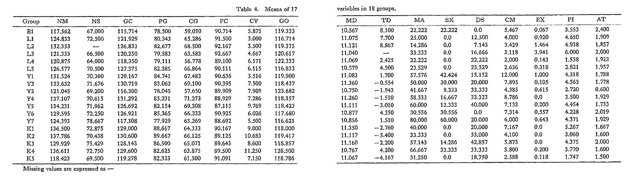
L5 is the last group of the Later Jomon population. Its 25 specimens belong to the Nagasaki University Collection, except for one from the Goryo site that belongs to the Kyushu University Collection. These were excavated at several shell middens widely distributed along the coast line of northwestern Kyushu, including the neighboring Goto Islands. In Plate 3, the Wakimisaki skull is shown as an example. It has a relatively large face with a slightly convex profile, well developed squarish and massive mandible with small gonial angle, and remarkable gonial eversion. Y1 is a Yayoi group from the sand dunes at the Doigahama and Yoshimo sites in Yamaguchi prefecture and the Koura site, Shimane prefecture, on the north and west coasts of western Honshu (Fig, 4C). These 33 skulls belong to the Kyushu University Collection. They comprise a very interesting population, on which discussions concerning immigrants from abroad have been focused. The Doigahama skull shown in Plate 4 exemplifies the series, with its very long face and straight profile, large mandible with dominant chin prominence, large gonial angle, and dominant gonial eversion. These characteristics of the group seem especially strong in the Koura skull shown in Plate 4.
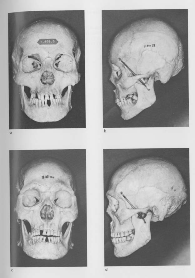
Y2 is from the northern part of Kyushu, These 19 skulls belong to the Kyushu University Collection. They were obtained from burial jars rather than from pit-graves in sand dunes, as were the Y1 group, at several sites in Fukuoka City and vicinity. As shown in Plate 5, the faces of this group are long when viewed frontally, as in the Y1 group. In the lateral view, however, alveolar prognathism is the most striking trait.
Y3 consists of 13 skulls from the Nagasaki University Collection. They were excavated from the sand dunes at Otomo, a small village in Saga prefecture fronting the Japan sea. The example of this group shown in Plate 5 has a relatively long face and slightly convex profile, with a small mandible accompanied by a large gonial angle and the still-evident trait of gonial eversion. This group has been considered an intermediate Yayoi type from northern and northwestern Kyushu, and has been a matter of interest and controversy for a long time. Naito (1981) concluded, after a comparative morphological study, that they do indeed belong to the northwestern Kyushu group. In the present investigation, however, this group is again separated from the northwestern Kyushu group, for the purpose of testing the differences between various samples from the standpoint of dental disease. Y4 consists of 14 specimens, partly from the Kyushu University Collection and partly from the Nagasaki University Collection. They were found in burial jars excavated at many sites in the Tsukushi Plain of western Kyushu. As seen from the Mitsu site example in Plate 6, their faces show a very similar pattern to that of the Doigahama people (Y1),but are larger than the latter and differ further in that the facial profiles are more convex. Another matter of interest in this group is that the facial bones were dyed with Indian red or a cinnabar-like substance.
Y5 consists of 15 specimens from the Nagasaki University Collection, They were excavated at the Futatsukayama site in the Tsukushi Plain. Groups Y4 and Y5 might properly be combined with each other, but they have been kept separate in the present study for the purpose of testing the peculiar characteristics of Y4, which seem to indicate a similarity with the Jomon people. From the facial pattern exemplified in Plate 6, differences between these two groups can be seen in the shorter facial dimension of the Y5 group, especially in the size of the mandible. Also, in the Y5 group, the facial prognathic trend is more prominent with a protruded maxilla. The facial bones are also dyed very slightly red, as in the Y4 group. Y6 is the largest group in the present study. It consists of 70 skulls from the Nagasaki University Collection. They were buried in different styles-in sand dunes, burial jars, and stone coffins-at many sites widely distributed along the coastline of northwestern Kyushu and in the neighboring islands. This group is well known in Japanese anthropology, and Naito (1971), after studying these materials, suggested that morphological differentiation was already present among the Japanese population in the Yayoi period. The example skull shown in Plate 7 was excavated at the Nejiko site. It shows a very short and squarish face, which is very similar to that of the Hegi people (L4) from the Later Jomon period, though it is much larger than the Hegi faces. The mandible is slender, but gonial eversion is evident.
Y7, the last group of the Yayoi population, consists of 14 specimens from the Kyushu University Collection. They were excavated from sand dunes at the Hirota site, Tanegashima Island, to the south of Kyushu. As seen in Plate 7, the vertical dimension of the face is relatively short and the profile line shows an alveolar prognathic trend. The mandible is not large, but is well developed, with a very small gonial angle and no evidence of gonial eversion. K1 is the first group from the Kofun period, obtained from the Kyushu University Collection. It consists of nine samples excavated at several burial mounds in the northern part of Kyushu. As shown in Plate 8, the skulls have long faces, narrow mandibles with slight gonial eversion, and prognathic profiles.
K2 consists of 10 specimens from the Kyushu University Collection, excavated from burial mounds in northeastern Kyushu (Fig. 4D). An example photograph in Plate 8 shows an extremely long face with a large mandible. A prognathic trend is also dominant. Gonial eversion is not evident. The facial bones were dyed red, as in the case of the Mitsu people (Y4).
K3 is made up of eight specimens from the Kyushu University Collection. They were excavated from several burial mounds in the northern Tsukushi Plain, which opens along the Chikugo River. The sample photograph in Plate 9 shows a relatively squarish face with a bialveolar protruded facial pattern and slight but evident gonial eversion. The facial bones were dyed red.
K4 consists of 10 specimens from the Kyushu University Collection, excavated from burial mounds in the southern half of the Tsukushi Plain. As shown in Plate 9, their faces are very long and show a dominantly prognathic pattern. K5 is the last group of the Kofun period, and of all the groups studied in the present investigation. It consists of 17 skulls from the Nagasaki University Collection. They were excavated from several burial mounds in the inland region of southern Kyushu. The sample photograph in Plate 10 shows a relatively small skull, almost the same in size as those of the El group. The facial plane angle is large, the angle of convexity is small, and alveolar protrusion is evident.
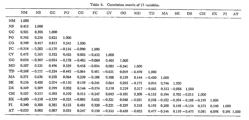
MethodsFor the purpose of analyzing differences and similarities between each pair of groups, 17 variables were selected to express the characteristics of all populations. As shown in Table 3, the first eight variables were obtained by means of roentgenographic cephalometrics to represent cranial and skeletal morphology.
The roentgenographic cephalometric device is an instrument for making accurate reproducible lateral roentgenograms of the head (Broadbent, 1932; Hofrath, 1932). The geometric conditions of this tool, namely the distance from the X-ray anode to the median sagittal plane of the skull, and the distance of the skull to the X-ray film, are kept constant (these are 150 cm and 15 cm, respectively, in the present study, in accord with current practice), so that it is possible to compare the linear and angular measurements among different individuals. Nasion-Mcnton (NM) and Nasion-Sclla (NS) were used to represent the vertical and antero-posterior dimensions of the facial cranium, respectively. Breadth of the cranium was not employed as a measurement in this study, as it ia naturally not available from lateral roentgenograms. Gnathion-Condylion (GC), Pogonion-Gonlon (PG), and Condylion-Gonion (CG) express the size of the mandible. The next two variables, facial plane angle (FC) and angle of convexity (CV), derive from angular measurements which correspond to the anteroposterior relationship of the middle and lower face, or facial prognathism. The eighth measurement, Gonial Angle (GO), relates to mandibular shape. Tooth crown modulus of the mandibular first molar (MD) was obtained by direct measurement of the mesio-distal and bucco-lingual dimensions of the tooth on the skulls, and used to represent tooth size. Total discrepancy (TD) is the actual size of the tooth-to-denture-base discrepancy, which is calculated from tooth size, size of dental arch, and forward inclination of mandibular incisor by use of a modified Tweed's method of orthodontic diagnosis (Tweed, 1944, 1945; Inoue et al,, 1971). Namely, the available arch length is measured on the mandible of the skull, from the mesial side of the first molar on one side to the same point of the opposite side. The measurement is taken along the dental arch by using soft, annealed brass wire. The required space is obtained by summing the mesio-distal crown dimensions of all the teeth from the central incisors to the second premolars on both sides of the mandible. Then the arch length discrepancy is obtained by subtracting the required space from the available arch length. Finally, the arch length discrepancy is modified by adding a correction factor which corresponds to the excess forward inclination of the mandibular incisors. This correction factor, the head plate correction, is obtained by dividing the difference between FMIA (the angle of the long axis of the mandibular central incisor to the Frankfort horizontal plane) and 65 degrees (Tweed's standard value of FMIA) by 1.25 (the constant for converting degrees into millimeters). The final value of the total discrepancy is the sum of the arch length discrepancy and the head plate correction. In these procedures for calculating the total discrepancy, a missing value for a tooth is replaced by the measured value of the same kind of tooth on the opposite side of the dental arch. When the same kinds of teeth on both sides of the dental arch, or any other necessary measurements such as FMIA, are missing they are replaced by the mean values of the corresponding items of the group to which the case belongs. The next three items relate to the type of occlusion. Usually, each malocclusion is diagnosed from a clinical standpoint and classified according to four categories: maxillary protrusion, anterior cross-bite, crowding, and bialveolar protrusion. But in the preseiit study, these factors are all added together and expressed as the prevalence of malocclusion (MA). There are four major pathogenic factors of malocclusion (Inoue et al., 1971), but only two of them are treated in this study. The skeletal factor (SK) is employed as one of the important variables in the present investigation, because it relates to the size and shape of the jaw bone (Hanihara et al., 1981), and is understood to have a special significance in the increase of malocclusions in the Yayoi period (Inoue et al; 1984), The prevalence of the discrepancy (DS) is a qualitative expression of the tooth-to-denturc-base discrepancy. It is the rate of the carriers of the symptoms, such as forward inclination of the incisors, crowding of the teeth, and protrusion of the alveoli, which are the inevitable consequences of the tooth-to-denture-base discrepancy. It is expected that this item will have a very close correlation to the total discrepancy (TD) in quantitative expression. Neither functional nor dental factors were employed in this study, for these factors relate mainly to the developing stage of occlusion. The last four variables relate to traits of cultural and pathological stress imprinted on teeth. They include the ratio of dental caries and missing teeth to present teeth (CM), ratio of teeth extracted by ethnic custom to present teeth (EX), extent of alveolar bone resorption possibly induced by periodontal diseases (PI), and extent of occlusal tooth attrition (AT). This latter relates to cooking and eating behavior (Begg, 1965), and shows the highest significant correlation with the passage of time among our dental pathological characteristics (Inoue, 1983). In the present study, all the necessary data are from previous reports of the authors (Inoue et al., 1982c, 1982d), in which the raw data obtained by the measurement of this material using the above-mentioned methods were shown. Among the variables used in this study, measured values which were expressed by proportional measures, namely all the roentgenographic cephalometric measures, tooth crown modulus, and total discrepancy, were simply processed to get the means and standard deviations for each group. Group means and standard deviations were also obtained for scores of periodontal index and tooth attrition, as well as the ratio of carious and missing teeth and the ratio of extracted teeth. Those items which had nominal measure ments and were expressed by prevalence (%) for each group, such as malocclusion, skeletal, and discrepancy factors, were arc sine converted before standardization. The means of these 17 variables for 18 cases are shown in Table 4; in Table 5 the data are standardized to place the mean values at 0.000 and the standard deviations at 1.000.
Using these standardized data, a factor analysis for 17 variables and a Q-mode principal components analysis for 18 groups were conducted using the SPSS batch system at the University Computer Center of the University of Tokyo. A cluster analysis for 18 groups was also employed, using a Q-mode correlation matrix. In order that the influence of morphological and dental pathological characteristics could be separately discussed, the same series of analyses was applied separately for the nine variables which express cranial and skeletal morphological characteristics (Nasion-Menton to tooth crown modulus of the mandibular first molar) and for the eight variables which represent dental pathological conditions or cultural influence (total discrepancy to occlusal tooth attrition). FindingsFactor AnalysesThe correlation matrix obtained by factor analysis for 17 variables is shown in Table 6, and the varimax rotated factor matrix with eigenvalues for each factor is shown in Table 7. Two dimensional expressions of variables are shown in Fig. 5.
As estimated from the factor loadings seen in Table 7 and Fig. 5, the first factor seems to relate to the general size of the facial skeleton and the teeth. This may be decided not only by the specific morphological characteristics of the groups, but also by the nutritional condition of their ecosystem. This factor may be termed the general size factor.
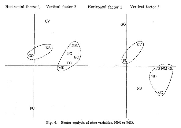
The second factor is probably controlled mainly by the occlusal type that relates to the skeletal factor. The effect of the distance Nasion-Sella is also dominant, probably because these two variables are linked; both are deviations in the aiitero-postcrior dimension. The ratio of carious and missing teeth and periodontal disease also contribute to this factor. Thus, this factor may be considered the occlusion-pathological factor. This factor seems to be influenced by the type of food and eating behavior during the individual's life time. Not only arc dental caries and periodontal disease predominantly affected by oral environmental pollution, but also the hypcrgrowth of the mandible is possibly related to the increased mastication induced by food characteristics. The third factor may be related to the tooth-to-denture-base discrepancy. This factor is also concerned with the antero-posterior relationship of the maxilla and mandible, or facial prognathism. Thus, it may be named the discrepancy-prognathism factor. It seems to be regulated by the cumulative effect of the foods eaten by an individual's ancestors over a long period of time. Tooth attrition behaves dependently with this factor. The fourth factor relates to the custom of tooth extraction, and again to periodontal disease and facial plane angle. The fifth factor relates to the gonial angle. However, the eigenvalues of these two factors are not large, and therefore they do not seem to be of particular significance. The above-stated understandings seem to agree with the distribution of the variables shown in Fig. 5, in which the variables are well separated by the general size, occlusionpathological, and discrepancy-prognathism factors. The results of the factor analysis for nine morphological characteristics, from NasionMenton to tooth crown modulus of the mandibular first molar (NM to MD), are shown in Table 8 and Fig. 6. Again, the dominant factor seems to be general size; it explains as much as 62.6% of the variance. The second and third factors are obviously those for prog nathism and the shape of the mandible, respectively. Figure 6 shows the interrelationships of these variables.
Table 9 and Fig. 7 show the results of the factor analysis for eight variables relating to occlusion and dental disorders. These range from total discrepancy to occlusal tooth attrition (TD to AT). In this case, variables obviously relating to the first factor are the prevalence of malocclusion which relates to the skeletal factor, and the ratio of carious and missing teeth. The second factor represents the tooth-to-denture-base discrepancy accom- panied by tooth attrition. The third factor represents a combination of periodontal disease and the custom of tooth extraction. These relationships are very well expressed in Fig.7.
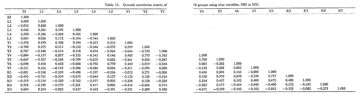
Q-mode Principal Components AnalysesTables 10, 11, and 12 show the Q-mode correlation matrices of 18 groups obtained by using 17 (NM to AT), nine (NM to MD), and eight (TD to AT) variables, respectively. Table 13 and Fig. 8 show the result of a Q-mode principal components analysis carried out using the correlation matrix in Table 10. From the results of the factor analysis and the distribution of the size factors seen in Table 4, the first principal component seems to relate to the general size factor; correspondingly, there seems to be a trend for groups with smaller crania to be on the left side of the analytical space in Fig. 8.
The second principal component is probably controlled by a complex including angle of convexity, prevalence of malocclusion, skeletal, and discrepancy factors. The third principal component has a close relationship with carious and missing teeth as well as periodontal disease. The fourth principal component seems to relate again to the malocclusion and skeletal factor. These two variables were probably acting here on the prevalence of malocclusion as caused by the skeletal factor, while in the second principal component they were working as determining elements of facial prognathism. The fifth principal component relates to many variables such as dental caries, extracted teeth, occlusal tooth attrition, and so on, and it does not seem to be very significant. As shown in Fig. 8 the groups representing the Later Jomon, Yayoi, and Kofun periods are formed by the first and second, and the first and third principal components, respectively. However, there are several inconsistencies:
Table 14 and Fig. 9 show the results of a Q-mode principal components analysis of nine variables, from Nasion-Menton to tooth crown modulus of the mandibular first molar. The purpose of this analysis was to study the effect of morphological characteristics in separating the groups. In this case, the first principal component is strongly related to Nasion-Menton, namely the vertical dimension of the face.
The second principal component relates to the angle of convexity and the facial plane angle, namely the facial prognathism factor. The third principal component relates to mandibular size; Gnathion-Condylion, Pogonion-Gonion, and Condylion-Gonion, as well as the tooth crown modulus of the mandibular first molar, all contribute. The fourth and fifth principal components relate to the mandibular shape and antero-posterior dimension of the face, respectively. A two-dimensional expression of the analyzed groups, using the first and second, and the first and third principal components, is shown in Fig. 9 on the left and right, respectively. Groups are formed which fit the various time periods, but they are spread over wide ranges and are not separate from each other. Table 15 and Fig. 10 show the results of a Q-mode principal components analysis which was carried out using eight variables, from total discrepancy to occlusal tooth attrition. This was done in order to investigate the effect of dental disorders in characterizing the prehistoric populations.
The first principal component seems to be controlled by the two variables of the toothto-denture-base discrepancy, namely, the total discrepancy and the prevalence of discrepancy. The second principal component relates to malocclusion, especially of the skeletal type. The third principal component is concerned with the dental diseases related to dental caries and periodontal disease. The fourth, and last, principal component seems to be governed by tooth attrition as well as by the prevalence of discrepancy as a modifying factor of tooth attrition (Inoue, 1982e). As shown in Fig. 10, separation of the groups by period is fairly good, though several groups-5, Y6, and K.4-re out of their proper areas on the left as is K1 on the right. Cluster AnalysesFigure 11 shows a single linkage dendrogram of 18 groups obtained by a cluster analysis run on the Q-mocle correlation matrix in Table 10. There are three major divisions; an isolate group consisting only of Y7; a larger group made up of K4, Y3, K5, and L4; and a much larger group which can be divided into three minor clusters and several isolates. The first minor cluster consists of three Later Jomon populations, L1, L2, and L3, which were excavated from shell middens in eastern Japan. The second minor cluster includes three Kofun period groups, K1, K2, and K3, which were excavated from burial mounds located close to each other in northern Kyushu. The third minor cluster consists of three Yayoi groups, Y2, Y4, and Y5, all from burial jars excavated in northern. Kyushu.
The three minor clusters have in common the facts that their component groups belong to the same period, and that the sites from which they were excavated are relatively close to each other. Except in these three cases, however, there are few commonalities in the periods and locations of the sites grouped together in the analysis. Figure 12 is another single linkage dendrogram of 18 groups, obtained by using nine morphological variables from Nasion-Menton to tooth crown modulus of the mandibular first molar. This dendrogram shows how the 18 groups cluster according to morphological characteristics.
Two major clusters and a very small intermediate cluster are shown in Fig. 12. One of these two clusters, at the upper part of the dendrogram, consists of only Yayoi and Kofun groups, but includes no Jomon groups. In the other main cluster, in the lower part of the dendrogram, the groups are mainly of the Earlier and Later Jomon and Yayoi periods, and only two Kofun populations are represented. It is also notable that within the main clusters, subclusters are formed on the basis of geographic proximity. For example, K1, K2, and K3, all from northern Kyushu, buildup a subcluster; Y2, Y4, and Y5 are in almost the saine relationship. Furthermore, L5 of the Later Jomon period and Y6 of the Yayoi period, which were excavated from almost the same area, formed a subcluster. Populations LI and L2 from the Later Jomon period and Y1 from the Yayoi period were all excavated from sites outside Kyushu, and they form a minor cluster. The remaining samples, El, Y3, K4, L4, L3, and K5, are spread temporally across the Earlier and Later Jomon, Yayoi, and Kofun periods, but nevertheless show a certain geographic commonality with each other, all being from peripheral parts of Kyushu. The dendrogram in Fig. 13 is derived from a cluster analysis using the Q-mode correlation matrix displayed in Table 12, which was constructed using eight variables relating to dental disorders. In this case, the similarities and differences between samples are more clearly controlled by time periods than by geographic neighborhoods. All the Later Jomon groups are clearly separated from the groups of other periods in a cluster covering a wide area from northern Honshu (LI) to northwestern Kyushu (L5). The Yayoi and Kofun groups seem to be relatively close to each other. Groups Y3, Y7, and K4 form one small cluster, and El and Y6 another. The remaining Yayoi and Kofun groups form a large cluster, in which they are separated incompletely.
DiscussionCertain trends in the distribution of the analytical groups with reference to time period and geographical clustering are of particular interest. They are treated immediately below. Inconsistencies observed during the analysis also require discussion, and they will be taken up in a following section. Similarities of Groups by Periods and Geographical DistributionIn Fig. 8 left, the Later Jomon groups are located on the lower left side of the vertical axis; this distribution is probably controlled by facial convexity and the tooth-to-denture-base discrepancy. The Early Jomon group is located to the far left, close to the horizontal axis of the diagram. In contrast, the Kofun populations lie on the right side of the vertical axis, distributed around the horizontal axis. The Yayoi groups are widely scattered, lying between the Jomon and Kofun groups above the horizontal axis. It is very interesting hat the Yayoi groups can be divided into two types by the general size factor. Groups Y3, Y6, and Y7 belong to the smaller type and are located on the left side of the vertical axis, while Y2, Y4, and Y5 are of the larger type distributed on the right side of the axis, overlapping with the Kofun population. Interestingly, the Y1 group, consisting of the Doigahama and Koura people who are conventionally called "tall immigrants," fall in to the smaller type. These two sets of Yayoi groups are well separated from each other. From these findings, it may be inferred that the general size of the facial skeleton has gradually enlarged through time, with time being expressed along the horizontal axis from left to right of the diagram. On this assumption, it is most likely that the four Yayoi groups of the smaller type, and K5 of the Kofun population, belong morphologically to an older type close to that of the aboriginal Jomon population. In the same manner, the three groups of the larger type, Y2, Y4, and Y5, are morphologically similar to the Kofun people and may be seen as representing a newer type of Yayoi population. These hypotheses agree with Naito's (1971) suggestion that several types of Yayoi people could be distinguished in early west Japan. In addition, it is very interesting that these three groups were all interred in burial jars, except for the Y6 people, who used modified burial jars in a few cases (The Board of Education, Arikawa Town, 1977). Thus, the groups studied in the present investigation may be divided into several families. The most dominant family formation observed is the Yayoi-Kofun population, in which groups Y2, Y4, Y5, K1, K2, and K3 are included. It is very interesting that all of these groups are from northeastern and northern Kyushu and the northern part of the Tsukushi Plain, a relatively narrow strip in northern Kyushu. Furthermore, those groups with red-dyed facial bones, Y4, Y5, K2, and K3, all belong to this family. The Y2 and K1 groups, both from northern Kyushu, are perhaps best understood as the most progressive groups of their respective periods, as far as the locations of these groups in Fig. 8 are concerned. The custom of tooth extraction is not observed in this family with very few exceptions in Y2, Y5, and K4, Gonial eversion is no longer dominant, the only exception being the slight expression of this trait in K3. The second family which may be identified is the Jomon-Yayoi population. It consists of L4, L5, Y1, Y3, Y6, Y7, and K5, which are from northwestern and northeastern Kyushu, inland southern Kyushu, and western Honshu; these groups have in common the fact that their native places are distributed around the periphery of northern Kyushu. The third group, minor in comparison to the first two, consists of L1, L2, and L3, which come from eastern Japan. These seem to comprise the oldest type of the Later Jomon population; they may represent the old Jomon population, or a Jomon population of east Japan type, because El, which is the sole group in the sample that is known to be from the Earlier Jomon period, does not seem to belong to this population. The El group may be a component of the Jomon-Yayoi population. Groups El and L4, both dwellers in the same mountain cavern during the Earlier and Later Jomon periods respectively, have no characteristic shared traits except the minimal tooth extraction. Comparable situations are also observed in Fig. 8 right, although here the Yayoi and the Kofun groups are better separated, probably because of the extremely high rate of dental disease in the Yayoi period. The dendrogram in Fig. 11 reflects the same conditions. In Fig. 9, which was obtained using only morphological characteristics as variables, the differences between periods are further reduced. The groups of each period are spread over wide and overlapping morphological ranges. By contrast, morphological characteristics are more firmly controlled geographically. Groups L1, L2, and L3, representing the Jomon population of the east Japan type, are located from top center to top left of the left-hand diagram. Groups Y1, Y6, Y7, L5, K5, L4, and Y3, the Jomon-Yayoi population, are distributed from the upper right to the lower left. Finally, groups Y2, Y4, Y5, K1, K3, and K4, the Yayoi-Kofun population, which seems to be the newer family, are distributed in the lower right half of the figure. However, note that the Jomon-Yayoi population, the older family, is spread out by the influence of mandibular shape in Fig. 9 right. The cluster analysis displayed in Fig. 12 is highly congruent with these findings. In Fig. 10, based on dental pathological factors, separation of the samples by time periods is clear in both the left and right figures. Though geographic factors are still reflected in a limited way in the left-hand figure, they are not significant in the right-hand figure. In Fig. 13, characteristics are predominantly grouped by period rather than by geographical region, except in the case of several peripheral groups. On the basis of these analyses, it appears that in the earlier stage of the period studied, there are fewer geographic differences among the populations. The groups tend to cluster more according to the period to which they belong. In the later stage, however, characteristics which are specific to the period are reduced, and similarities and differences among the groups are influenced more by geography. It may also be inferred that geographic differences and similarities among the groups are defined by morphological characteristics, and that the difference between each period relate strongly to dental pathology. In other words, morphological characteristics of groups are controlled by their geographic environment, while dental-pathological properties of groups are governed by the culture of the particular period. These facts suggest that culture can spread rapidly over a wide area, but morphological characteristics are highly stable, corresponding with geographic conditions. Inherited physical characters can be altered only through admixture, which requires a long time to bring about changes. There is another reason why morphological measures reflect the characteristics of the geographic environment. This is because human morphological size is strongly influenced by nutritional conditions, which in turn depends on the productiveness of the ecosystem. As for morphological size, Hiramoto (1972, 1977) showed that the estimated stature of the Japanese male stayed within the range 1591.1 mm-1630.6 mm, with a fluctuation rate of 2.5%, throughout the millennia from the Later Jomon to the Kofun period. This means that size variations among the populations were relatively small when compared with those of dental diseases. On the other hand, the mean stature of 20 year old Japanese boys has increased 42.0 mm (2.5%) in the last 13 years (Ministry of Health and Welfare, Japan, 1983), suggesting that morphological size can easily change as a result of an alteration in nutritional environment. Because of these two characteristics, i.e., a relatively small variance among groups and adaptability to environmental stress, there may be some limitations to be considered in employing morphological size as a measure for comparing groups which are located close to each other. This opinion seems to agree with that of Hanihara (1981), who recommends the use of non-metric characteristics, and Omoto (1981), who employs genetic analysis in his research. As for dental pathology, it is quite natural that dental disorders should express the characteristics of cultural periods. These periods are not morphologically, but archae-ologically defined, using cultural vestiges as the measure, and dental disorders are reflective of human culture. They are imprinted on the teeth and on the patterns of occlusion through the reduction of jaw bones and the pollution of the oral environment as a result of cultural practices of food selection and preparation. Inconsistencies in Group PatterningThere are several inconsistencies in the location of Yayoi and Kofun groups within the analytical spaces of Figs. 8-13, though the Jomon placements seem to be stable and show no major contradictions to expectations about their proper temporal and geographical positioning. The first problem to be addressed under the present heading concerns group Y3, which was excavated at the Otomo site, Saga prefecture. It was separated from the northwestern Kyushu group (Y6) in this study, for the purpose of testing the degree of similarity between the two groups. Y3 is located at the upper left part of the Yayoi circle in Fig. 8 left, but in the right figure it deviates toward the lower left direction, far beyond the other Yayoi groups. This may be because the Y3 specimens have much less dental disease than the other Yayoi populations. The morphological characteristics of Y3 are more clearly expressed in Fig. 9 left and right, in which the Y3 group is located at the leftmost position close to the Later Jomon groups. However, it stays at a quite reasonable position among the Yayoi populations in Fig. 10. These facts may indicate that group Y3, the Otomo people, belongs morphologically to the older type of Yayoi population or to the Jomon-Yayoi population, though the people had already adopted Yayoi culture. As for the differences and similarities between Y3 and Y6, a Yayoi group from northwestern Kyushu, both of them belong to the Jomon-Yayoi population, showing very close values of general size and malocclusions. However, they show also minor differences in prognathism, dental caries, the vertical dimension of the face, the mandibular shape, and the tooth-to-denture-base discrepancy. From these facts, it may be inferred that both groups are descendants of the aboriginal Jomon people and developed distinct characteristics when isolated from each other. A second point of concern is the relationship between Y4 and Y5. As mentioned before, these two Yayoi groups were excavated from almost the same area in the Tsukushi Plain and there is no reason for separating them from each other except to examine a certain peculiarity of the Y4 (Mitsu) people. The Mitsu people are of special interest. Comparing the Yayoi groups, Kanaseki et al. (1960) pointed out a strong physical similarity between the Mitsu and Doigahama (Y1) people. They also suggested similarities between the Doigahama people and the Kofun people, as well as with the Jomon people from Tsugumo (L3). Naito (1971), after studying a large number of samples of Yayoi populations from northwestern Kyushu (represented by Y6 in this study), presented his opinion that the northwestern Kyushu population is different from the Doigahama and Mitsu people, but similar to the Jomon people from Tsugumo. In the present study, there are some very close relationships in morphological and dental pathological characteristics between the Y1 and Y6 populations, and between Y4 and the Kofun population. Moderate similarity between L3 and Y6 was also confirmed for morphological characteristics. Limited morphological similarities between the Y1-Y4 pair, and between Y1 and the Kofun groups, as well as dental pathological similarity between L3 and the Kofun groups, were observed. The other pairs of groups, Y1 and L3, Y4 and L3, and Y4 and Y6, show no evidence of close relationship. Thus, it may be that the Y1 and Y6 groups, both of which belong to the old-type Yayoi population, have similar morphological as well as dental pathological characteristics. On the other hand, Y4 should belong to a newer type of the Yayoi population, and as far as the results of the present study are concerned, it is quite likely to belong to a transitive stage from the Yayoi to the Kofun period. As shown in Figs. 8-13, the Y4 and Y5 groups are located very close to each other with no dominant inconsistency. Thus the peculiar characteristics of the Mitsu people could be understood as a proper expression of the unmixed characteristics of the newer type Yayoi group, or the Yayoi-Kofun population. Y7 from Tanegashima Island is also one of the older type Yayoi groups. It is to be expected that there was very little human and cultural interchange between the northern part of Kyushu and Tanegashima Island; this is undoubtedly the reason for the retention of old characteristics in the island population. The K4 and K5 groups were excavated from peripheral areas of the Kyushu district, namely the southern part of the Tsukushi Plain and the inland zone of southern Kyushu. These groups are located very close to the Jomon circle in Figs. 8-10, as if they could be classified as Jomon-Yayoi populations. In other words, the Kofun people can also be divided into two types, both by morphology and dental pathology. Finally, it is very important to make reference to the immigrants from abroad who might have brought rice agriculture. Kanaseki et al. (1960) suggested that there was an inflow of immigrants who differed from the aboriginal Jomon people in physical characteristics. On the basis of the extremely high rate of skeletal factors of malocclusion found for the Yayoi period, the present authors have also pointed out that the Yayoi people might include immigrants from abroad (Inoue, et al., 1984). However, the results of the present study seem to suggest that none of the specific groups examined here are an immigrant population. In other words, the immigrants probably did not come in all at once as a large group, but instead as small groups entering occasionally, mainly into the northern part of Kyushu. This was the center of Japanese culture at that time, spanning the periods of the older and newer Yayoi populations. Thus the Jomon-Yayoi populations, including K4 and K5 in the present study, may comprise the aboriginal groups present in Japan before the inflow of the immigrants, or just at the time of the influx. On the other hand, the Yayoi-Kofun population represents the new Japanese after admixture had progressed. These conclusions agree essentially with those of Naito (1981) who emphasized the compatibility of Kanaseki's Influx Theory and Suzuki's Transformation Theory of Japanese population history. Concerning the time of introduction of rice agriculture in the Later Jomon period, many authors have presented their opinions (Sahara, 1969; Sakura, 1964; Turner, 1979). Most of them seem positively or passively to agree that rice was introduced into Japan before the Yayoi period, These opinions accord with the present discussion, in that the inflow of immigrants is seen as having already begun in pre-Ynyoi times, during the Later Jomon period. ConclusionsThis paper has addressed some possibilities for using dental pathology as a measure to evaluate similarities and differences between prehistoric populations. The results show that dental diseases, which are strongly influenced by human culture, are an appropriate tool for the development of new perspectives on the prehistory of Japan. The conclusions may be summarized as follows. 1. In the earlier prehistoric ages, the time factor was the main determinant of similarities among the populations studied, but in the later period, the geographic factor became predominant. 2. Morphological factors relate strongly to the geographic distribution of groups because admixture changes morphology only slowly, and because of the ready adaptability of morphology to the environment. By contrast, dental pathological factors represent characteristics determined through the influence of culture, and they are subject to more rapid change. 3. The Yayoi population may be rationally divided into two morphological types, an older and smaller Jomon-Yayoi type and a newer and larger Yayoi-Kofun type. In other words, the population that developed from the Jomon period to the Kofun period might be divided into the aboriginal Jomon, the Jomon-Yayoi, and the Yayoi-Kofun families. 4. Most of the inconsistencies seen among the Yayoi group seem to result from a thought process in which an effort is made to divide two morphological groups into three archaeological periods. a) The northwestern Kyushu Yayoi group (Y6) and the Otomo group (Y3) belong to the same Jomon-Yayoi population morphologically, but represent different levels of culture in terms of dental disease effects. b) Both of the Tsukushi Plain Yayoi groups (Y4 and Y5) belong to the newer Yayoi-Kofun type morphologically and they are different from the Doigahama-Koura group (Y1) as well as from the northwestern Kyushu Yayoi group (Y6), both morphologically and dental-pathologically. c) The Doigahama-Koura group (Y1) and the northwestern Kyushu Yayoi group show considerable similarities with each other, morphologically as well as dental-pathologically. 5. As for the immigrants who are believed to have introduced rice agriculture to Japan, they do not seem to have come all at once as a large group, but rather to have come as small groups from time to time, mainly into the northern part of Kyushu. Their arrival apparently coincided with the transition between the older and newer Yayoi populations. These conclusions include several important points requiring further examination. In the future it will be necessary to improve methods of analysis and the selection of variables to obtain increasingly reliable results. Such studies should extend the area from Japan to all of Asia, and ultimately to the world as a whole, and expand the period of concern to include the Medieval and current ages. AcknowledgementsThe authors wish to express their sincere appreciation to Professors Kazuro Hanihara, Department of Anthropology, Faculty of Science, The University of Tokyo; Masafumi Nagai, Department of Anatomy, Faculty of Medicine, Kyushu University; and Yoshiatsu Naito, Department of Anatomy, Faculty of Medicine, Nagasaki University, for their kind permission to investigate their respective collections of Japanese skeletal remains. The authors would also like to extend their thanks to Associate Professor Takeru Akazawa of the Department of Anthropology and Prehistory, The University Museum, The University of Tokyo, and Professor C. Melvin Aikens of the Department of Anthropology, University of Oregon, for kindly inviting us to participate in the Japanese prehistory symposium of the XIth International Congress of Anthropological and Ethnological Sciences, where this paper was originally presented. We are also indebted to Dr. Kiyotaka Koizumi, Department of Anthropology, Faculty of Science, The University of Tokyo, for his kind advice and assistance in data processing. References
|