DESCRIPTION OF INDIVIDUALS
Minatogawa 1
In the upper jaw, UI2, UC and UP2 of the right side, and UI2, UM1, UM2 and UM3 of the left side are preserved, while in the lower jaw, full dentition except for the incisors and the right LC is preserved.
Since all the teeth are affected by heavy attrition, detailed observation of the crown characteristics is hardly possible.
UI2, UC and UP2 of the right side and UI2 of the left side have lost more than two-thirds of the crowns by attrition. In the left UM1, more than two-thirds of the crown has been lost by attrition, and tooth wear almost reaches to the tooth neck at the lingual side. As a result, the occlusal surface shows a steep slope from the buccal side down to the lingual side.
Also in the left UM2, about a quarter of the crown at the buccal side and almost three-quarters at the lingual side are worn down. The occlusal surface thus shows a relatively steep slope, if not so steep as in UM1.
The left UM3 is relatively small in size, and shows a reduced type of crown shape, although the crown is affected by relatively heavy attrition. The crown probably is the "3+" type denned by Dahlberg (1949) because a small projection of enamel at the distal side suggests the presence of a reduced hypocone. The slope of the occlusal surface caused by attrition is weak in comparison with UM1 and UM2.
In the lower jaw, heavy attrition again affects all the teeth preserved, so that a detailed description of the crown shape is almost impossible. In contrast to the upper molars, the occlusal surface in the lower molars is nearly horizontal. Interproximal attrition is advanced to a considerable extent in both upper and lower molars.
The size of the crowns seems relatively small in the incisors, canines and premolars compared with the molars in both jaws. The size sequence of the molars is, judging from the crown modules, M2>M1>M3 in the upper jaw and M1>M2>M3 in the lower jaw. In both jaws, size reduction in M3 is relatively advanced, and this is partic ularly the case in UM3 which shows the "3+" type of crown mentioned above. In contrast to this, the crown of LM3 is probably five-cusped, with a remarkably large protoconid.
Scott (1979a) proposed a new technique to evaluate degrees of attrition which allows more highly precise scoring than the method of Broca. According to this scoring tech nique, the degrees of attrition in the molars are counted as follows: UM1 =38, UM2= 34, UM3=23; right LM1=31, LM2=22, LM3 =20; left LM1=33, LM2=22, and LM3 =20. The differences between Ml and M2 are 4 points in the upper molars, and 9 and 11 points in the lower right and left molars, respectively. These differences, especially those in the lower molars, are considerably larger than those in most modern populations. For instance, Scott (1979b), who studied Amerindian skulls, reported mean differences of 1.57-0.47 points for the upper jaws and 1.30-0.03 points for the lower jaws. According to this author, the larger difference indicates heavy attrition from a young age. From this point of view, it is likely that tooth attrition advanced quite rapidly in this individual, and this seems to suggest that the Minatogawa man ate a large quantity of coarse or tough foods.

Minatogawa Mandible A
This is an isolated mandible, probably of a female. The teeth preserved are LC, LP1, LP2, LM1 and LM2 of the right side, and LP2, LM1 and LM2 of the left side. Judging from the degrees of attrition, this individual seems to be younger than the Minatogawa 1 and Mandible C. LM3 is congenitally missing on both sides.
The right LC lost about a quarter of the crown by attrition and dentin is exposed at the tip of the cusp. On the lingual surface, the mesial marginal ridge and the central ridge are well developed, and the lingual tubercle is also large. In the right LP1 dentin is exposed at the base of the protoconid, but the enamel still covers the lingual side of the occlusal surface. The metaconid seems to be small.
In the right LP2, the degree of attrition is about the same as in the right LP1, and the dentin is exposed only at the buccal side of the occlusal surface.
In the right LM1, attrition shows Broca's 3rd stage, and all the cusps are worn down to make the occlusal surface nearly flat. Judging from the contour of the crown, this tooth is probably five-cusped with a relatively small hypoconulid. The groove pattern is not necessarily apparent but may be assumed to be Y-shaped.
The degree of attrition of the right LM2 is Broca's 2nd stage, small dentin exposures being seen at the areas of protoconid, metaconid and entoconid. The crown appears to be four-cusped without a hypoconulid, and the groove pattern is plus-shaped.
In the teeth on the left side, LP2, LM1 and LM2, attrition is relatively weak com pared with those on the right so that the crown shape is somewhat apparent. LP2 shows almost the same morphological condition as the right LP2 with the dentin exposed mainly at the buccal side of the occlusal surface.
LM1 is probably five-cusped, and the groove pattern shows a Y-shape. Attrition is much less than that of the right LM1 but the occlusal surface is flat.
The occlusal surface of LM2 is nearly flat, but, as in LM1, attrition is somewhat less than the right LM2. The crown is probably five-cusped with a small hypoconulid. The groove pattern is Y-shaped but is very close to a plus-pattern.
According to Scott's scoring method, the degrees of attrition are as follows: right LM1=26, right LM2=17, left LM1=19, and left LM2=17. As described above, the degrees of attrition are generally weaker in the left side teeth than in the right.
A special mention should be made of the tooth extraction of LI1 on both sides of this mandible. The sockets for these teeth are completely closed, and the alveolar processes form a sharp concave ridge. It is quite difficult to judge whether the extractions were made artificially or not, but the following points may suggest that this is an example of artificial tooth extraction:
1) LI1 on both sides were extracted symmetrically;
2) All the remaining teeth remained in quite healthy condition, and the condition of the tooth sockets suggests that they carried healthy teeth, although both LI2, left LC and LP1 are missing;
3) The condition of closure of the tooth sockets for LI1 suggests that both teeth were extracted at almost the same time, because the alveolar processes were absorbed symmetrically to make an almost complete arc.
If this is really an example of artificial tooth extraction, it is quite possible that it is the earliest instance known in Japan. The oldest examples so far reported are those from the early and middle Jomon Age (ca. 4,000-5,000 years B.P.), and the sites where they were found are scattered in the western part of Japan. In addition, those early cases show artificial extraction of LI1 of both sides, the same pattern as in the present case (T. Akazawa, personal communication).
In Okinawa, on the other hand, no such case has been so far reported from the Jomon Age, but only from the Yayoi period (ca. 2,000 years B.P.). In China, cases of artificial tooth extraction were reported from the Neolithic age of about 5,000 years before present, which is almost the same age as the Jomon Age. For this reason, the tooth extractions in the present case attract our particular attention.

Minatogawa Mandible B
Five right mandibular teeth, LC through LM2, are implanted in a fragment of man dibular corpus. The crowns are heavily worn down and the occlusal surfaces are nearly flat in all the teeth preserved.
Although only about one-third of the lingual surface of LC remains, it is supposed to be relatively simple in morphology with a very small lingual tubercle and very weakly developed marginal ridges. The occlusal surfaces of LP1 and LP2 are oval in shape.
Detailed observation is hardly possible because of heavy attrition, but the crown of LM1 is probably five-cusped judging from its contour, and that of LM2 seems to be four-cusped without the hypoconulid.
The scores of attrition by Scott's technique are 29 in LM1 and 21 in LM2. The difference between LM1 and LM2 is relatively large, as it is in the other individuals from the Minatogawa site.
The size sequence of the molar crowns is LM1> LM2, crown modules being 12.05 and 11.45, respectively.
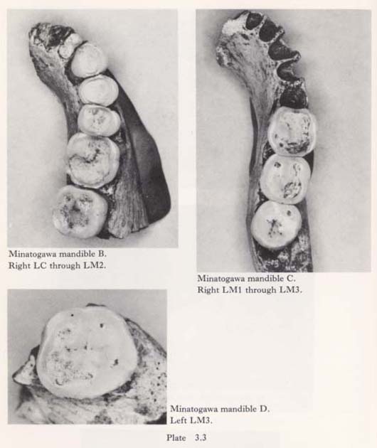
Minatogawa Mandible C
This individual is thought to be a female. The teeth preserved are LM1, LM2 and LM3 of the right side. As in the Minatogawa 1, heavy attrition does not allow detailed description of the crown morphology.
The dentin is fully exposed in the right LM1, partly in the right LM2 and only at the protoconid area in the right LM3. The degrees of attrition are expressed using Scott's scoring technique as follows: right LM1=30, LM2=22 and LM3=17. As in the case of Minatogawa 1, the difference of scores between LM1 and LM2 is very large, and this fact also seems to mean that attrition advanced more rapidly in this individual than in modern man. Interproximal attrition is remarkable in the three molars.
The molar size sequence is M1>M2>M3, but the size of LM3 is relatively large compared with the same tooth in Minatogawa 1.
Minatogawa Mandible D
This is a small fragment of the mandibular corpus with the left LM3. The dentin is exposed at the distal half of the crown, and the occlusal surface is almost flat.
Judged from the contour of the crown, this tooth seems to carry five main cusps, No interproximal attrition is observed.
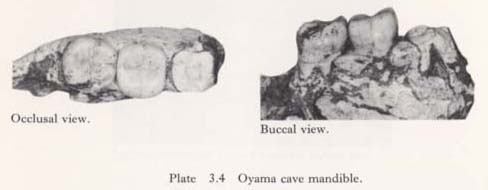
Oyama Cave Mandible
Only two molars, right LM2 and LM3, are implanted in a fragment of mandibular corpus, the crown of LM1 being broken off and the roots remaining in the tooth socket. This individual is relatively young because tooth attrition is not as advanced and the enamel still covers the occlusal surface completely.
The morphology of the occlusal surface is simple, with no accessory tubercle to be particularly mentioned. The groove pattern is a plus-shape in LM2 and a Y-shape in LM3. No hypoconulid is observed in LM2, but there is a trace of this cusp in LM3.
Swelling of the buccal surface is relatively strong, and the buccal groove in LM3 is divided into two branches at the tip, suggesting the presence of a weakly developed protostylid.
The size of the crown is considerably larger in LM3 than in LM2, the crown modules being 12.2 and 11.5, respectively. Degrees of attrition scored by Scott's technique are 12 in LM2 and 8 in LM3.
COMPARISON OF CROWN SIZE
Generally, the size can be divided into two components, size component and shape component. In the present study these two components will be compared separately between Minatogawa man and some other early and modern hominids.
The Minatogawa man used in this analysis is Minatogawa 1, a male who carries the largest number of teeth among the six individuals reported above. The teeth used in this comparison are UI2, UC, UP2 through UM3, and LC through LM3, and the measurements used are mesiodistal crown diameters.
The hominid groups compared are 18 populations, among whom are modern Japanese who have been measured by Hanihara, one of the present authors, and Jomon people, mainly from the middle and later stages of the Jomon Age, measured by Ueda, also one of the present authors. The data for modern Chinese, Aleuts, Javanese and Swedes are those compiled by Moorrees (1957), and data for the remaining populations, mainly fossils, are those compiled by Wolpoff (1971).
To compare the size component of the tooth crown diameters, the first principal component scores, which represent the size factor, were used (Tables 3. 2 and 3. 3).


As a general trend, it is quite evident that tooth crown size diminishes from earlier to later types of hominids. The largest tooth crowns are carried by australopithecines, and the next largest by Homo erectus. Among Homo sapiens, Neandertals are significantly larger in tooth size than the sapiens man.
It is quite interesting that Minatogawa 1 carries relatively large teeth among sapiens man, very close to that of the Upper Paleolithic man. The crown size of the Minatogawa 1 is even somewhat larger than that of the Pecos Pueblo, whose teeth are ranked as among of the largest in recent populations. The teeth of Minatogawa 1 are remarkably larger than those of modern Japanese and Jomon people, who show relatively small teeth in spite of their early existence (Neolithic; ca. 3,000-4,000 years B.P.). On the other hand, the upper molars of Minatogawa 1 are somewhat smaller than those of the Niah Cave man from the Upper Pleistocene (Brothwell, 1960), and much smaller than those of Australian Aborigines (Wailbri; Hanihara, 1976), Talgai (Campbell, 1925) and Keilor (Adam, 1943).
At any rate, it is worthy of note that the teeth in Minatogawa 1 are larger than those in most of the modern populations compared, and the difference is particularly remarkable between this individual and the Jomon people as well as modern Japanese.
For comparison of the shape component, Q-mode correlation coefficients, which represent only differences in the shape component, were employed. The populations compared are the same as those used for comparison of the size component.
As shown in Table 3. 4, the Minatogawa 1 is very close to Wadi Halfa of the Mesolithic age and Australopithecus robustus. This might be caused by a larger size difference between the molars and the remaining teeth. Bushmen and Pecos Pueblo are also relatively close to Minatogawa 1, but modern Japanese, Swedes and Jomon people are a little different from them. On the other hand, Minatogawa 1 is farthest from Homo erectus, and considerably distant from Neandertals whose incisors are proportionately larger than those of the other hominids. In general, analysis of the shape component clearly shows that Minatogawa 1 has relatively large molars in comparison with its incisors, canines and premolars.
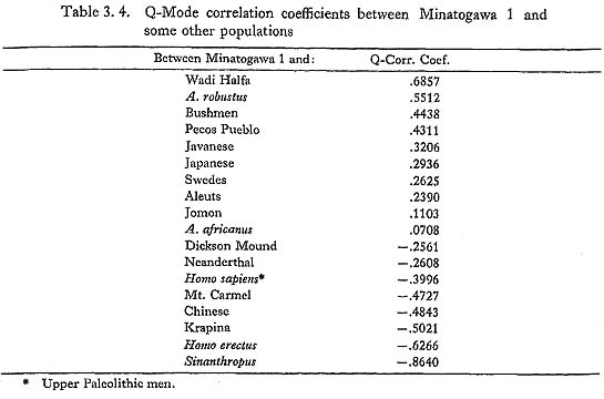
Differences in relative size of tooth crowns are also represented by some principal components, of which the second principal component seems best to show differences between molars and other teeth (Tables 3. 2 and 3. 3). On the basis of these scores, Minatogawa 1 is again very close to Australopithecus robustus, and very far from Homo erectus and Neandertals. Many types of recent man are relatively close to Minatogawa 1 in this respect.
Overall, an analysis of tooth crown size appears to show that the teeth of Minatogawa 1 are larger than those of most modern populations; in this sense, it may still retain an archaic characteristic, at least so far as the size component is concerned. In regard to shape, however, it is quite evident that the size difference between the molars and the remaining teeth is larger than most of the hominid groups compared.
This result does not necessarily show the evolutionary status of this individual, but it may be safely stated that Minatogawa 1 shows an intermediate tooth size betweenearly hominids who retain larger tooth size and recent hominids with somewhat smaller teeth. In other words, the size of the incisors, canines and premolars has reduced to a considerable extent, but the molars still remain relatively large.
SUMMARY OF DENTAL MORPHOLOGY
Although heavy attrition does not allow detailed description, it is quite likely that the crown morphology of the Minatogawa man is relatively simple, and this is particularly evident in the lower molars implanted in the Minatogawa Mandible A, in which attrition is less advanced. At the same time, the hypocone in the upper molars and the hypoconulid in the lower molars are generally weakly or not at all developed. The groove pattern in the lower molars is probably Y-shaped in LM1, but fully or nearly plus-shaped in LM2.
The reduction of the third molar is also apparent not only in morphology but also in crown size. The molar size sequence is dominantly M1>M2>M3 with a few exceptions which show the sequence M2>M1.
The difference in attrition between Ml and M2 is, as far as can be judged by Scott's method scores, relatively larger in comparison with modern man. This fact may suggest that their chewing force was very powerful.
In regard to crown size, difference between the molars and the remaining teeth is apparent, the former being proportionately much larger than the latter.
The size component of tooth crown measurements shows that the Minatogawa man belongs to a group of sapiens man with large teeth. In particular, their teeth are much larger than those of modern Japanese and Jomon people from the middle and later stages.
On the other hand, the shape component shows a somewhat different picture. The large size difference between the molars and the other teeth may suggest that the Minatogawa man is at the intermediate stage between early hominids with larger teeth and later hominids with smaller teeth. As stated above, the former is represented by the molars and the latter by the remaining teeth.
As a whole, the teeth of the Minatogawa man show mixed characteristics of modern and archaic hominids, the modern characteristics being mainly represented by crown morphology and molar size sequence, and the archaic characteristics by crown size or by the size component in particular.
Finally, special mention should be made of probable artificial tooth extraction in Mandible A. The tooth sockets for LI1 of both sides are completely closed, forming a sharp concave ridge; the reasons why this closure suggests artificial tooth extraction have already been described.
It is naturally quite difficult to decide whether the cause of such tooth loss was artificial or pathological, but it is still possible that this is a typical example of artificial tooth extraction which prevailed in early ages. If this individual really belongs to the Upper Pleistocene, this case is undoubtedly the earliest example found in Japan or, in fact, in all of Asia.
REFERENCES
- Adam, W. (1943)
- The Keilor fossil skull: Palate and upper dental arch, Mem. Nat. Mus., Melbourne, 13:71-77.
- Brothwell, D. R. (1960)
- Upper Pleistocene human skull from Niah Caves, Sarawak. Sarawak Mus. J., 9; 323-349.
- Campbell, T. D. (1925)
- Dentition and Palate of Australian Aboriginal. Univ. of Adelaide.
- Dahlberg, A. A. (1949)
- The dentition of American Indian. W. S. Laughlin (ed.), The Physical Anthropology of the American Indian, pp. 138-176, Viking Fund Inc.
- Hanihara, K. (1976)
- Statistical and Comparative Studies of the Australian Aboriginal Dentition. The Univ. Mus., The Univ. of Tokyo, Bulletin No. 11.
- Moorrces, C. F, A, (1957)
- The Aleut Dentition. Harvard Univ. Press.
- Scott, E. C. (1979a)
- Dental wear scoring technique. Am. J. Phys. Anthrop., 51: 213-218.
- Scott, E. C. (1979b)
- Principal axis analysis of dental attrition data. Am. J. Phys. Anthrop., 51:203-212.
- Wolpoff, M. H. (1971)
- Metric Trends in Hominid Dental Evolution. The Press of Case Western Reserve Univ.
|