PART I. SEASONAL DATING METHOD
II. Shell Structure and Growth Increments of Meretrix lusoria (Röding)
II-1. Growth Increments and Growth LinesThe terminology for shell growth increments and periodicities was reviewed by Rosenberg (1975): an increment is a repetitive unit of structure or composition in the skeleton, and the boundary is the structural or chemical limit of the increment or series. Hence, the growth line used in the present study is defined as an increment boundary that appears as a narrow light stripe when observed in thin section under the microscope, or as a narrow stripe of etchant-resisting material. According to Rosen berg (1975), one unit of growth increment spans the beginning of a rapid calcification portion to the end of a slow calcification portion, or the growth line. In other words, a growth line is the light portion of an increment which consists of dark and light bands in a thin section. If a growth line forms once a day, then the unit added during a daily cycle will be the daily increment. Additionally, the width of a daily increment is the thickness of the interval between daily growth lines, or the amount of deposit formed within a daily period. II-2. Methods of ObservationFor the observation of growth lines under an optical microscope, the shells were first embedded in polyester resin and cut along the line connecting the umbo and the midpoint of shell length at the ventral margin. The cut surface was then polished mechanically with carborundom sheets of different grade, and occasionally further with aluminum, and etched with an aqueous solution of 0.38% HCl for 5-10 minutes at room temperature. Replicas of the etched surface were made using 0.034mm thick cellulose acetate film (Obiken, bioden film). The replicas were observed with an optical microscope at a magnification of 40 to 400. Shell structure was studied by viewing thin sections of the shells through a polarizing microscope. A scanning electron microscope (SEM; Hitachi-HSM-2) was used to observe the finer details of the growth increments. Two kinds of etchant were used for SEM. To observe the relationship between crossed lamellae and growth lines, a mild etchant, 0.1 mol EDTA-4% paraformaldehyde solution was applied for 30-60 seconds. On the other hand, a solution of 0.38% HCl applied for about 3 minutes etches the ground surface so severely that the crossed lamellae structure becomes very faint, although the growth lines remain clear. II-3. Microstructure of Growth IncrementsThe Meretrix lusoria shell consists basically of an inner and an outer layer (Koike, 1973). In the outer layer, the reflected increments with a series of semicircular growth lines are seen. The growth increments are thickest at the surface of maximum growth which lies roughly along the middle of the thickness of the shell viewed in radial-section. The right and left valves close tightly at the edge. In central radial sections (Fig. II-1-A), the growth lines outside the closing surface curve around the surface of maxi mum growth, while those inside the closing surface are rather straight, slanting almost parallel to the inner surface of the shell. That is, the stratification of the growth line changes from the reflected form to the slanting form at the closing surface. This closing surface divides the outer layer into an outer zone and an inner zone.
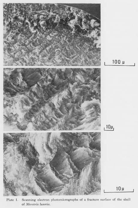
The outer layer of this species has a crossed lamellar structure (Kobayashi, 1967, 1971; Pannella and MacClintock, 1968). The lamellae radiate from the surface of maxi mum growth toward the outer and inner shell surfaces and intersect the growth lines when viewed in radial section under low magnification. The lamellar structure is made up of three orders of lamellae (Fig. II-1-B). Rectangular rods (third order lamellae) are arranged parallel to each other to form broad tablets (second order lamellae), which are stacked against each other to form first order lamellae. The rods of two adjoining first order lamellae incline in opposite directions, crossing at about 45-50 degrees. SEM photomicrographs of fracture surfaces of M. lusoria (Plate 1) show that the crossed lamellae observed under the optical microscope are first order lamellae. These lamellae are 5- to 10-µm-wide rows composed of thinner lamellae thought to be second order lamellae: which lie at opposing angles in neighboring first order lamellae.
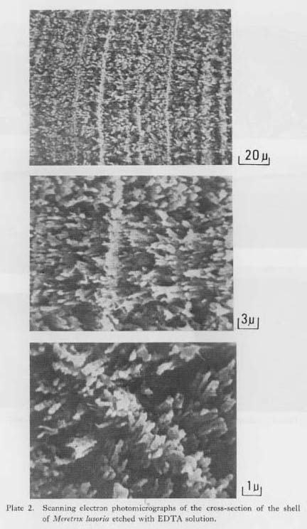
SEM photomicrographs of a radial section etched slightly with EDTA solution (Plate 2) show that the third order lamellae are etched into rod-like forms, and that the growth lines could be recognized as rows where the rods are especially dense. SEM microphotographs of surfaces etched with HCl solution (Koike, 1973: Plate 1) show growth lines as fine, bright striations on a dark background. The profile of a SEM image shows that growth lines are raised stripes of shell matter left by HCl etching, and that the dark background is a surface of material easily dissolved by the etchant.
In combining the SEM observations on the broken surfaces and those on the etched surfaces, the structural relationship between the crossed lamellae and the growth line can be illustrated as in Fig. II-1-B. The growth lines seen as growth bands under high magnification intersect the first order lamellae. A series of second order lamellae lie at opposite angles to each other, and growth lines run at an angle to the second order lamellae. The growth band represents a three-dimensionally-formed growth surface. Hence, the study of shell structure in relation to the growth line is of great interest in relation to microscopic shell growth or calcification. II-4. DiscussionIn order to apply growth-line analysis to seasonal dating of shells, distinctive features of the growth lines can be used to facilitate the counting of lines. A crossed lamellar structure is generally good, accompanied by clear growth bands (Pannella, 1975). The outer layer of the Meretrix lusoria shell has finer first order lamellae of the crossed lamellar structure than do those of Cardium (Cerastoderma) edule and Tapes (Ruditapes) aponica. The former has smooth and conspicuous growth lines, while the latter has discontinuous and zig-zagging lines. Moreover, the reflected type of growth lines in a crossed lamellar structure enables accurate measurement of growth increment widths along the surface of maximum growth (see Chapter IV-1). The surface of maximum growth of M. lusoria is easily determined from the orientation of the crossed lamellae. Growth lines are usually interpreted as organic-rich lines, and the etchant-soluble zone is a calcified layer (Pannella, 1975) based on SEM photomicrographs of the etched surface of Cardium edule in which growth lines were observed to be convex stripes. Although when etched with HCl solution, the growth lines of M. lusoria appear as convex stripes similar to those observed on C. edule, the studies in this paper suggest that growth lines are zones of high calcium concentration produced by microstructural differences, especially as seen in the density of crystal rods. High-magnification pictures of radial-section surfaces of M. lusoria etched with the milder etchant EDTA show that growth lines are zones composed of rods of third order lamellae similar to adjacent zones of the growth increment, but rods in the growth lines are aligned in greater density than in the adjacent zone. In a preliminary investigation using an X-ray microanalyzer, line-scanning patterns through growth lines and other growth increment zones indicated that the growth lines are a Ca-rich and Na-poor precipitate, suggesting that growth lines must be the dense part of rods of CaCO3. |