INTRODUCTION
Excavation of the Douara Cave in the 1970 field season uncovered certain quantities of the "amorphous silica," the microscopic structures of which seemed to be consistent with the silica skeletons isolated from some dicotyledonous plants though the plant names do not corresponded (Matsutani, 1973).
Many soil samples were also collected in the 1974 season and the author was able to engage in the study of these samples again.
This time a column sample was available as well as several ash samples.
Similar structures were also searched for among the spodograms of living plants con taining silicate which might have grown in the past in that part of Western Asia.
In addition, small fragments of charcoal were taken from an ash sample. Thin sections of these were made using a plastic resin and compared with modern samples of the probable equivalent.
MATERIALS AND METHODS
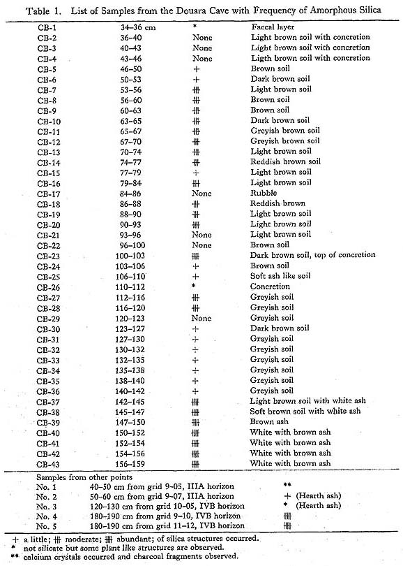
CB series of the column samples (Plate 1), each collecting 2 to 4 cm3 at the point X=9, Y=5.5, amounts to 43 samples. Five samples were collected from various ash layers as listed in Table 1. All these were processed according to the following procedure.

|
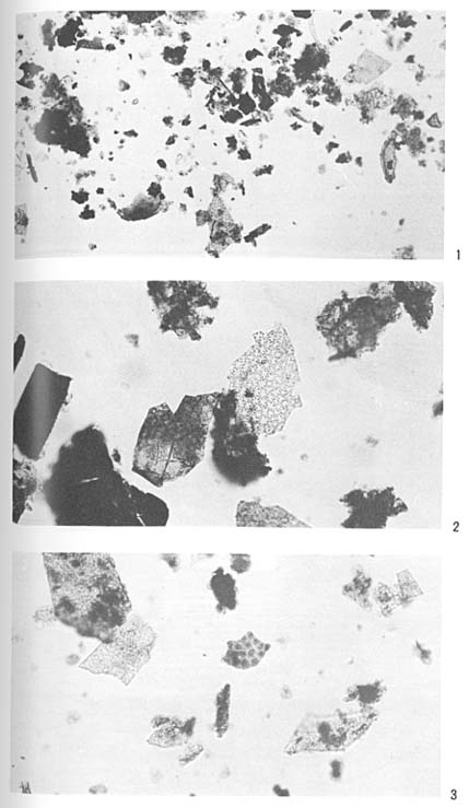
Photomicrographs of amorphous silica from the Douara Cave samples Fig. 2. No. 5(× 200). Fig. 3. CB-42(×200). |

Five random grams of each sample were transferred to a test tube, then 10 ml or more of 10% HCL solution was added to dissolve the calcium salts. Repeated washings with dis tilled water followed. The sample was then desiccated in an oven and mounted in Eukitt. They were stained with safranin or methylene blue in accord with the character of the silica skeletons.
Observations were made under a light microscope with simple polarized accessories.
RESULTS OF THE MICROSCOPY
In many of the samples mentioned above more or less identical structures as seen in the 1970 season were again observed. Their frequencies are shown in Table 1.
Regarding the CB-series shown in Table 1, the frequencies of the structures in question seems to have the following tendency.
The samples of the upper layers from CB-1 to CB-4 do not produce those structures; those from CB-5 to CB-20 except CB-17 show certain amounts of them. None occurred in CB-21-22; after that, up to CB-36 except both for CB-26 and -29, smaller quantities oc curred. Then again there was an increase and in the lower layers from CB-37 to CB-43, and the frequencies in these were the most abundant throughout all samples.
The other ash samples of various points, and the two from the greyish ash samples in the IVB horizon showed an abundance of these structures.
These suggest that the lowest horizon, IVB, associated with the Middle Paleolithic, yields these structures most abundantly. In order to determine their sources, spodograms of living plants were conducted and examined as described later.
In one of the ash samples regarded as to belonging to Horizon I, not silica structures, but calcium crystals were observed. This suggests derivation from some common leaf or wood. Among the ash small fragments of charcoal were found. Part of these incinerated fragments showed similar calcium crystals, and thin sections were made of these samples to determine the characteristics of the ash.
SPODOGRAMS OF THE SEEDS OF THE BORAGINACEAE
Many dicotyledons have been reported to contain silica in the epidermis of their leaves (Kohl, 1889; Netolitzky, 1912a, 1929; Geis, 1973 etc.), but few have been reported with regard to the seed coat or pericarp.
Failing to match the spodograms of plants collected around the cave which serve as food plants of the present day, other plants were investigated which might fit the require ments such as containing silica and growing in the areas of Western Asia. Borage seeds were selected and examined in the seed collection of the Botanical Garden, the Univer sity of Tokyo and in the herbarium specimens of the Department of Botany, the University Museum, the University of Tokyo.
The seeds from twenty three species within nine genera were incinerated in a porcelain dish by means of the dry ashing method using a muffle furnace at 550 C and were mounted in Eukitt after treatment with 10% HCL solution followed by washing and desiccating.
Results are listed in Table 2.
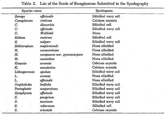
- Of the species examined, the following species have spodograms of wavy and thick cell membrane similar to the amorphous silica structures:
- Borago officinalis, Echium vulgare, Symphytum officinale, Symphytum peregrium, and Pentaglottis sempervirens (Plate 3).
However, these are not all the species of a given family, and the leaved portions were not tested because of a want of time and materials. Silica skeletons of hairs from the family, or skeletons of stomata were not found in the Douara samples, seeds being more probable than leaves. However, other families not examined might reveal similar structures.
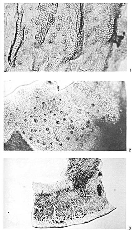
Photomicrographs of the spodograms of seeds of Boraginaceae Fig. 2. Symphytum peregrium (× 200). Fig. 3. Pentaglottis sempervirens (×200). |
PREPARATION OF THE CHARCOAL FRAGMENTS
Identification of the charcoal fragments with archeological connections attracted many workers with various approaches as summarized by Paulssen (1964) and Gannon (1973).
Charcoal fragments derived from the ash of the Douara Cave at the point described above were of such small size, about 0.7 mm long and 1.5 mm in diameter, that preparation by thin sectioning was conducted.
Techniques of embedding in plastic resin have been highly developed for use with elec tron microscopy, and epoxy resin has been successfully utilized for archaeological wood and charcoal samples (Gannon, 1973). However, here was used glycol methacrylate (GMA) which is missible with water and was recently developed for light microscopy in place of the conventional paraffin wax.
- The procedure of embedding the samples which follows mainly refers to Leduc and Bernard (1967) and Cole and Sykes (1974) with slight modifications.
- (i) The fragments were immersed in distilled water in glass vials until they sank to the bottom due to the penetration of water into the porous structures of the samples.
- (ii) Transferred to a solution of 50% solution of GMA with water, the samples were sub jected to a partial vacuum for around ten minutes.
- (iii) The vial was transferred to a rotator for good penetration and to avoid uneven polimerization.
- (iv) Samples were transferred to an 80% GMA solution and subjected the rotator.
- (v) Samples were immersed in pure GMA resin and rotated.
- (vi) Subjected to a resin mixture of GMA 7: n-butyl methacrylate 3 with 0.5% Benzoil Peroxide as an accelerator, the samples were again placed in the rotator.
- (vii) A prepolimerized mixture was prepared. The samples were transferred
to this and rotated.
Each step took approximately 24 hours or more. - (viii) Final polimerization in aluminum foil was conducted with ultraviolet light for five hours and was followed by desiccation in a 70°C oven.
Sectioning of this embedded specimen after the removal of the aluminum foil was done using an ultra microtome and a glass knife. About 2 thick sections were put on water drops with the aid of a syringe on the slide glass. After desiccation on a hot plate, the samples were mounted with immersion oil to facilitate the microscopy.
CHARACTERISTICS OF THE STRUCTURES
Sections of the charcoal fragments revealed under the microscope have the following traits as shown in Plate 4.
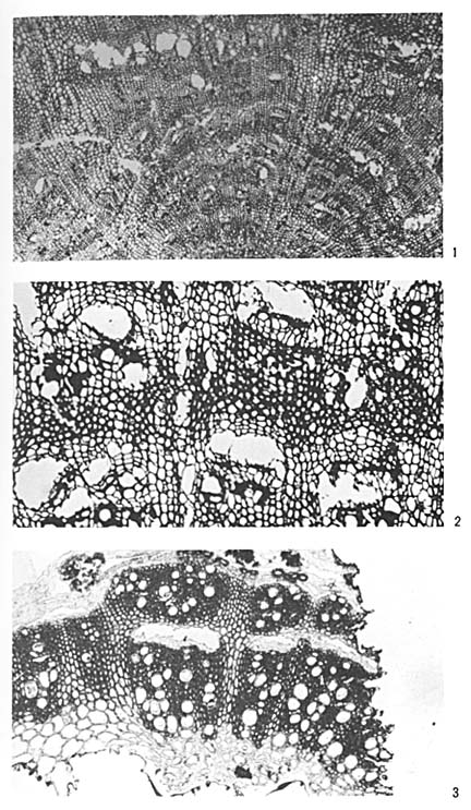
Fig. 2. Charcoal fragment (×200). Fig. 3. Stem of Chenopodiun cf. album. |
Groups of vessels accompanying included phloem are adjacent with each other and broad rows ofparechyma cells bind the vessel groups. Phloems are arranged in a concentric pattern.
These traits are characteristic of such plant groups of Centrospermae as Chenopodiaceae, Amaranthacease, and Nyctaginaceae (Balfour, 1965; Philipson and Ward, 1971, etc.).
Seen from Plate 4-1, the diameter of the cross plane is not large. This seemed to be not derived from a large tree, but from a slender twig of a young tree or a stem of herba-ceous plant.
According to the pollen analysis of the Douara Cave (Sohma and Sasaki, 1973), the pollen of Chenopodiaceae are well established and it is highly possible that the origin of the charcoal and ash, the latter revealing many small calcium crystals under the polarized microscope, are derived from the stems and/or leaves of chenopods. According to the report of the pollen analysis described above, five species of Chenopodiaceae, i.e., Halocnemum strobilaceum, Salsola villosa, Noaea mucronata, Haloxylon articulatum and Atriplex leucoclada, are listed as growing around the Palmyra Oasis.
Among the plants collected near the cave by a member of the 1974 field season, four specimens were identified as Chenopodiaceae by Mr. Ohba and GMA sectioning was carried out on these specimens.
GMA SECTIONS OF LIVING PLANTS
The specimens are preserved at the Department of Botany, the University
Museum, the University of Tokyo. Each slender stem about 2 to 3 mm in diameter
was cut to 8 mm thick and embedded in the resin by almost the same procedure
as was used with the charcoal fragments. However, presoftening was carried
out with haut-clave and the process began with a 20% solution followed by
30% and 40% solutions of resin.
Of the four samples sectioned for comparison, almost similar structures with those obtained from the charcoal were found only in one specimen identified as Chenopodium cf. album.
CONCLUSION
Microscopic study of one of the ash samples from the upper layer (Horizon
I) reveals that the ash comes from a plant which contained much calcium salts,
not silica, and that the charcoal fragments from among the ash are possibly
carbonized stems of Chenopodium.
Ash from the lowest layer (Horizon IVB) contained much amorphous silica; its micro-scopic structure resembles spodograms of seeds of some Boraginaceae.
DISCUSSION
It might sound strange that the Douara people of the Middle Paleolithic
might utilize the seeds of Boraginaceae, for the family is not to the best
of our knowledge on a familiar economic plant. The majority of the reports
with paleoethnological information are con-cerned with food plants such as
cereals and pulses; the borage family is seldom mentioned.
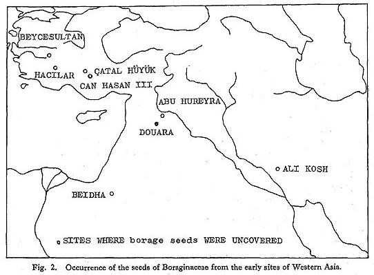
But in lists of plant remains from aceramic Hacilar in Turkey (Helbaek,
1970) and from the lowest level of All Kosh in Iran (Helbaek, 1969), however,
species of this family such as Heliotropim sp., Heliotropium persicum, Lithospermum
arvense, and Echium sp. were re-corded. Further, Dr. Helbaek notes the occurrence
of the seeds of this family from the early seventh millennium Beidha in Jordan
and from early Catal Hüyük in Turkey. He has com-mented (Helbaek,
1970:237) on these occurrences: "For some reason hitherto unexplained the
fruits of several species of this family occur with a conspicuous frequency
in very early deposits of plants, although the fruits can be of no nutritive
value whatever and the stems less profitable than most other."
It should be noted that both in the case of Ali Kosh and Hacilar their occurrence
diminished as time went on (loc. cit.).
Also, in the list of the plant remains of Abu Hureyra in Syria (Hillman,
1975) and Can Hasan III in Turkey (French, Payne and Hillman, 1972), Lithospermum
arvense, Echium sp., and Cf. Lithospermum arvense were found from the metholithic
and early neolithic period, but Dr. Hillman made no comment on these finds
which are not microscopic but macroscopic in nature. Dr. Netolitzky, who pioneered
spodograms in the field of archeological plant remains, related the detection
of Borago officinalis from the corpses of prehis-toric Egyptian mummies with
the aid of silicified membranes (Netolitzky, 1912a). Unfor-tunately, detailed
descriptions were not included in his report, so it is not certain whether
he was referring to the silica skeletons of leaves or seeds, although millet
and sedge accom-panied by borage were reported in detail in his other report
(Netolitzky, 1912b), Yet, we know from this that the ancient Egyptian people
utilized this species as a foodstuff.
It is highly probable, therefore, that people of Western Asia much valued Boraginaceae, and this might also be said of the Douara people.
Acknowledgments
The author would like to express appreciation to the following persons who have made this short report possible.
Firstly I extend my thanks to the members of the Tokyo University Scientific Expedition to Western Asia, especially to Mr. K. Endo, Department of Applied Sciences, College of Humanities and Sciences, Nihon University, and to Mr. T. Akazawa, National Science Mu-seum for sample collecting and information concerning them. Dr. H. Kanai of the National Science Museum suggested the Boraginaceae and Prof. Y. Furusawa, Ryukyu University, formerly of the Botanical Gardens, the University of Tokyo, gave permission to obtain seed samples.
To Dr. M. Wada, Department of Botany, Faculty of Sciences, the University of Tokyo, and to Dr. Y. Fukuda, Department of Biology, Faculty of General Education, Chiba Uni-versity, the author owes much for instructions and technical support during the making of the thin sections of charcoal fragments and on the identification of the sections of carbon-ized and fresh specimens respectively.
Mr. H. Ohba, Botanical Gardens, the University of Tokyo, kindly identified living plants collected around the cave.
To Professor N. Watanabe, Department of Anthropology, Faculty of Sciences, the Uni-versity of Tokyo, who led me to this field of spodograms and thereafter has encouraged me, the author owes much.
LITERATURE CITED
- Balbour, E. (1965)
- Anomalous secondary thickening in Chenopodiaceae, Nyctaginaceae and Amaranthaceae. Phytomorphology, 15: 111-122.
- Cole, M.B. Jr. and Sykes, S.M. (1974)
- Glycol methacrylate in light microscopy: a routine method for embedding and sectioning animal tissues. Stain Technology, 49: 387-400.
- French, D.H., Payne, R.J. and Hillman, G.C. (1972)
- Excavations at Can Hasan III 1969-1970.In: Papers in Economic Prehistory (E.S. Higgs, ed.), Cambridge: 181-190.
- Geis, J.W. (1973)
- Biogenic silica in selected species of deciduous angiospermums. Soil Science, 116: 113-131.
- Helbaek, H. (1969)
- Plant collecting, dry-farming, and irrigation agriculture in prehistoric Deh Luran. In: Prehistory and human ecology of the Deh Luran Plain (F. Hole et al., eds.), Memoirs of the Museum of Anthropology, University of Michigan, 1: 383-426.
- Helbaek, H. (1970)
- The plant husbandry of Hacilar. In: Excavations at Hacilar (J. Mellart, ed.), Edinburgh: 189-224.
- Hillman, G. (1975)
- The plant remains from Tell Abu Hureyra: a preliminary report. Proc.Prehistoric Society, 41: 71-73.
- Kohl, F.G. (1889)
- Anatomisch-Physiologische Untersuchung der Kalksaize und Kieselsäure in der Pflanze. Marburg.
- Leduc, E.H. and Bernhard, W. (1967)
- Recent modifications of the glycol methacrylate embedding procedure. J. Ultrastmct. Res., 19: 196-199.
- Matsutani, A. (1973)
- Microscopic study on the 'amorphous silica' in sediments from the Douara Cave. In: The Palaeolithic Site at Douara Cave in Syria, Part 1 (H. Suzuki and F. Takai, eds.):127-131.
- Netolitzky, F. (1912a)
- Kieselmembranen der dicotyledonenblätter Mitteleuropas. Öst. Bot.Zeitschr., 62: 353-359, 407-411, 466-473.
- Netolitzky, F. (1912b)
- Hirse und Cyperus aus dem prähistorischen Ägypten. Beih. z. Bot.Centrbl, 29,Abt.II: 1-11.
- Netolitzky, F. (1929)
- Die Kieselkorper. Linsbauer's Handbuch der Pflanzenanatomie, Berlin.
- Paulssen, L.M. (1964)
- Identification of active charcoals and wood charcoals, Trondheim: 1-113.
- Philipson, W.R., Ward, J.M. and Butterfield, B.G. (1971)
- Anomalous cambia, The Vascular Cambium-its development and activity, London: 95-110.
- Smith F.H. and Gannon, B.L. (1973)
- Sectioning of charcoals and dry ancient woods. Ameri-can Antiquity, 38: 468-472.
- Sohma, K. and Sasaki, M. (1973)
- Palynological studies on the deposits of the Douara Cave. In: The Palaeolithic Site at Douara Cave in Syria, Part 2 (H. Suzuki and F. Takai, eds.): 133-141.