Materials and Methods
|
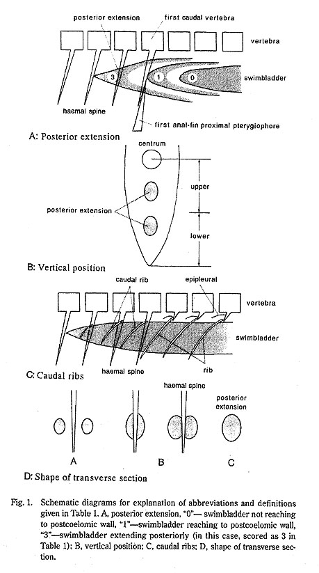
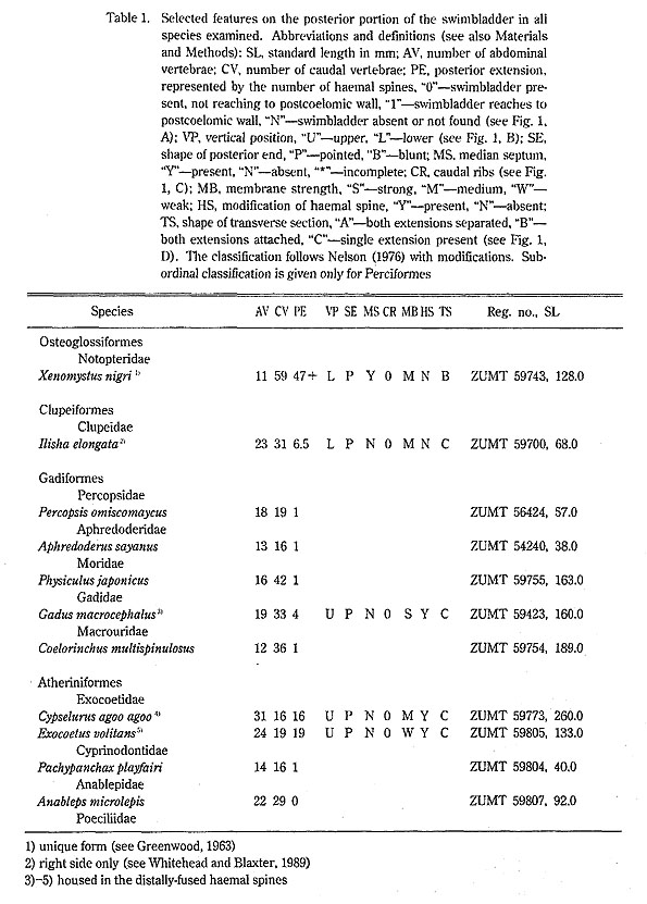
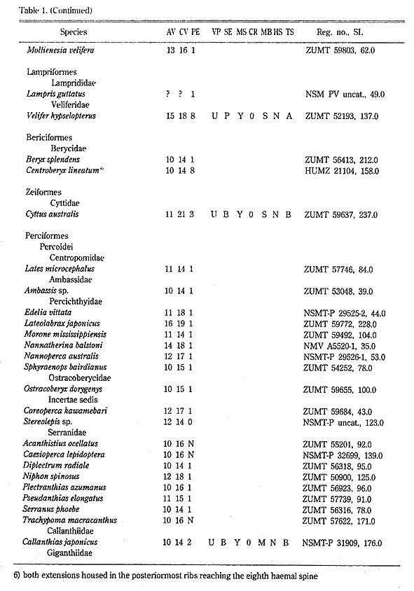
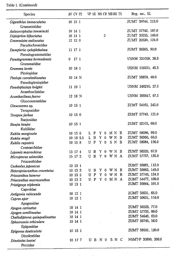
Although limits of the Percoidei differ between authors (Greenwood et al., 1966; Nelson, 1976,1984,1994; Johnson, 1984,1993). the familial classification of percoids used here follows Nelson (1976) with modifications. Specimens representing about 300 percoid and other teleostean species were dissected. Details of the posterior portion of the swimbladder were examined under a Wild M-8 binocular microscope after alizarin red S staining. X-ray negatives were used for vertebral counts. Serial (or a single) transverse sections of the body were prepared to allow more precise observations: 2-5 mm thick sections were cut from semi-frozen specimens and imbedded in commercial agar, placed in a shallow plastic container, and preserved in alcohol. The following aspects of the swimbladder were noted. 1) Posterior extension (Fig.1, A): The posterior part of the swimbladder extending beyond the first haemal spine and/or the first anal-fin proximal pterygiophore, into the myotomes of the caudal region of the body. The degree of extension is indicated by the number of the haemal spine associated with or just above the posteriormost part of the swimbladder. The extension of the swimbladder to the origin of the anal fin (e. g., Sciaenidae) or into the hollow space of the first anal-fin proximal pterygiophore (e. g., Lateolabrax of Percichthyidae) is not recognized as a posterior extension. 2) Vertical position (Fig.1, B): The position of the posterior extension was classified as either upper or lower at a vertical through in the middle part of the posterior extension. 3) Shape of posterior end: The posterior end is either pointed or blunt, although differentiation between the two may be relatively subjective. 4) Median septum: Usually either present or absent in species having a swimbladder with posterior extensions attached to either side of the haemal spines. An incomplete septum, however, was found in some species (e. g., embiotocids). Species with two extensions separated from the haemal spines are tentatively considered as having a median septum. "N" denotes species possessing a swimbladder with a single posterior extension. 5) Caudal ribs (Fig.1, C): Those ribs originating from the caudal vertebrae, not being serially-positioned epipleurals as found in some species. They usually support the posterior extension of the swimbladder. The caudal ribs identified in this study corresponded to "epihemal ribs" of Stiassny and Jensen (1987), but not of Morris (1982). 6) Membrane strength: Swimbladder membranes were classified as either strong, medium, or weak. 7) Modification of haemal spine: Applicable only when such modification was clearly or apparently associated with the posterior extension of the swimbladder. 8) Shape of transverse section (Fig.1, D): Taken at the middle portion of the swimbladder extension, three basic structures were recognized; two diverticula separated from each other ("A"); two diverticula attached to each other ("B"); a single diverticulum ("C"). Poll (1969) recognized three conditions (median, asymmetrical, paired) of the posterior extension of the teleost swimbladder. His "median" and "asymmetrical" categories are included in "C", and "paired" in either "A" or "B" in the present study.
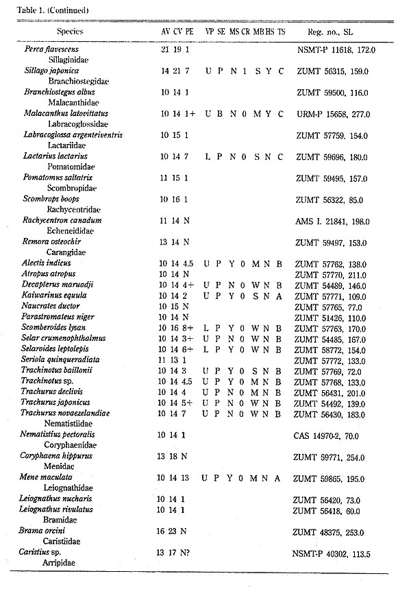
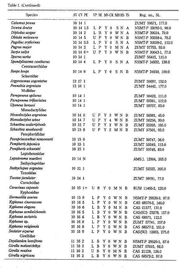
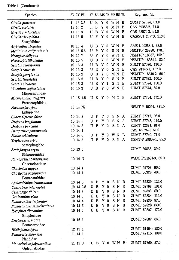
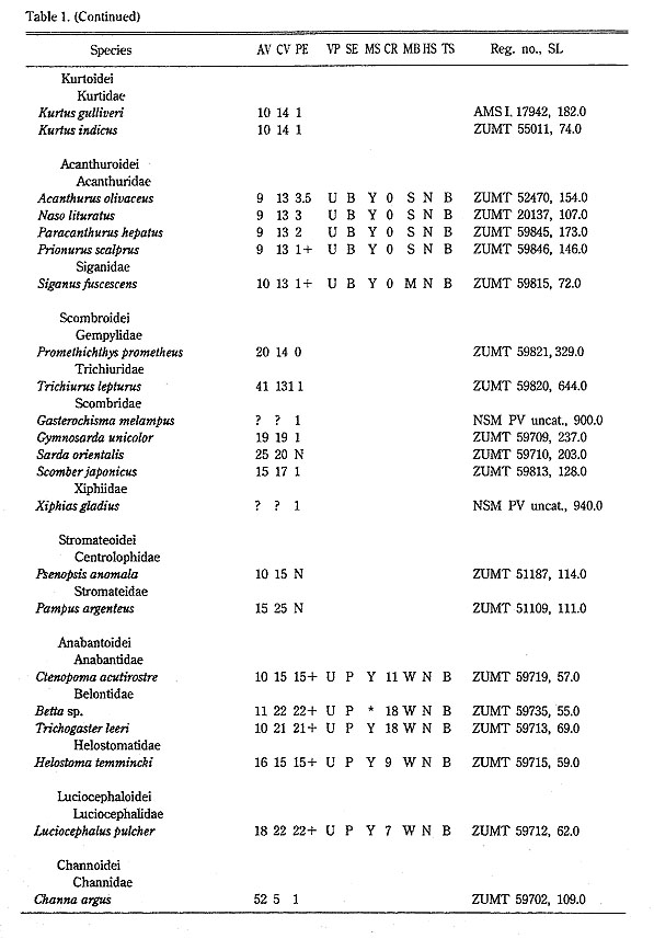
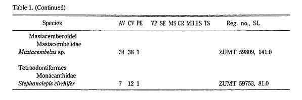
Study materials with standard length are listed in Table 1. Institutional acronyms follow Leviton et al.(1985).
In May 1994, while we were preparing the manuscript, Y. Tominaga untimely passed away. Although close to completion at that time, some sections required subsequent revision and rewriting by K. Sakamoto and K. Matsuura. |